. 2013 May 17;8(5):e62310.
doi: 10.1371/journal.pone.0062310. Print 2013.

Hypoxia stimulates the EMT of gastric cancer cells through autocrine TGFβ signaling

Affiliations

Hypoxia stimulates the EMT of gastric cancer cells through autocrine TGFβ signaling

Junko Matsuoka et al. PLoS One. .

Abstract

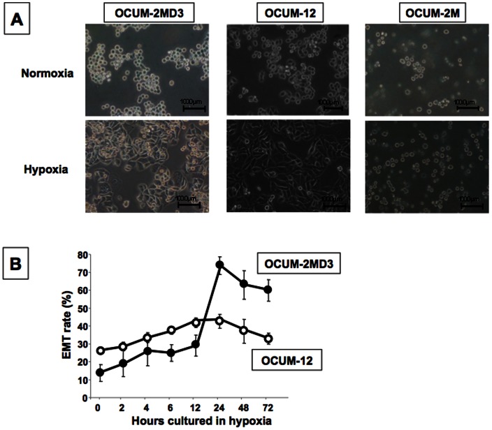
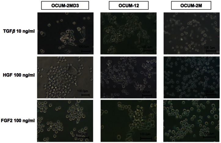
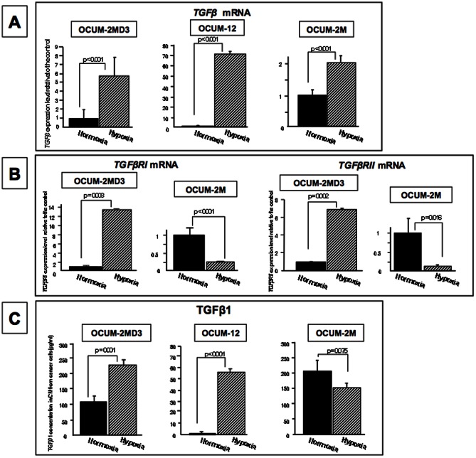
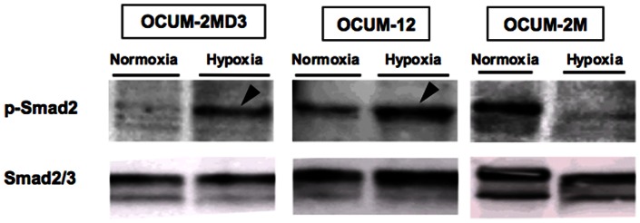
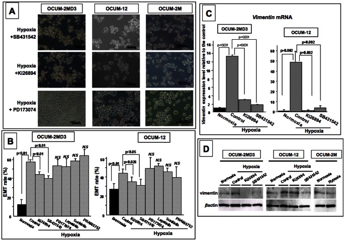
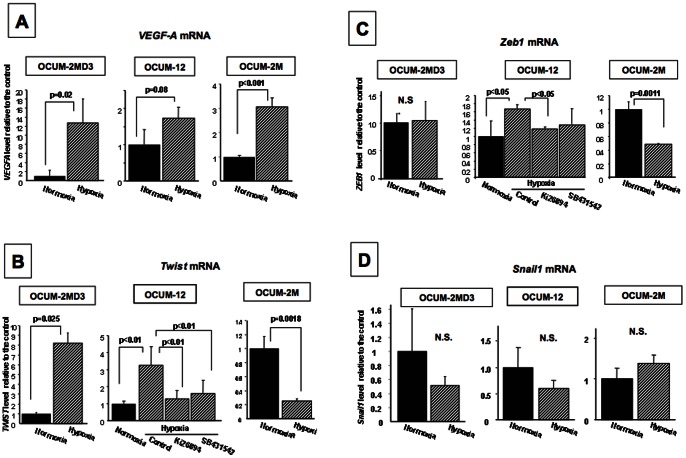
Epithelial mesenchymal transition (EMT) is considered to be correlated with malignancy of cancer cells and responsible for cancer invasion and metastasis. We previously reported that distant metastasis was associated with hypoxia in gastric cancer. We therefore investigated the effect of hypoxic condition on EMT of gastric cancer cells. Gastric cancer cells were cultured in normoxia (21% O2) or hypoxia (1% O2) for 24 h. EMT was evaluated as the percentage of spindle-shaped cells in total cells. Effect of transforming growth factor β1 (TGFβ1) or tyrosine kinase inhibitors on the EMT was evaluated. The expression level of TGFβ1 and TGFβR was evaluated by real time RT-PCR. The TGFβ1 production from cancer cells was measured by ELISA. Hypoxia stimulated EMT of OCUM-2MD3 and OCUM-12 cells, but not that of OCUM-2M cells. The expression level of TGFβ1 mRNA under hypoxia was significantly higher than that under normoxia in all of three cell lines. The expression level of TGFβR mRNA was significantly increased by hypoxia in OCUM-2MD3 cells, but not in OCUM-2M cells. TGFβR inhibitor, SB431542 or Ki26894, significantly suppressed EMT of OCUM-2MD3 and OCUM-12. TGFβ1 production from OCUM-2MD3 and OCUM-12 cells was significantly increased under hypoxia in comparison with that under normoxia. These findings might suggest that hypoxia stimulates the EMT of gastric cancer cells via autocrine TGFβ/TGFβR signaling.


Conflict of interest statement

Competing Interests: The authors have the following interests. KS, TS, and AM are employees of Kyowa Hakko Kirin Co. Ltd. Ki26894, which was used in this study, is a product of Kyowa Hakko Kirin Co. Ltd. There are no further patents, products in development or marketed products to declare. This does not alter the authors' adherence to all the PLOS ONE policies on sharing data and materials, as detailed online in the guide for authors.

Figures

Figure 1
Figure 1. Morphologic changes of gastric cells under a hypoxic condition.
(A) An increase of polygonal or spindle-shape cells was recognized in OCUM-2MD3 and OCUM-12 cells under hypoxia, but not in OCUM-2M cells. (B) The morphology of OCUM-2MD3 and OCUM-12 cells began to change after 4 h in hypoxic culture. EMT of OCUM-2MD3 and OCUM-12 cells was most evident at 24 h of culture under hypoxia. EMT was determined when the polygonal or spindle shape was found in cancer cells by phase-contrast microscope.
Figure 2
Figure 2. Effects of various growth factors on cancer cell morphology.
Morphologic change was found in the presence of TGFβ and HGF at 24 h of culture in OCUM-2MD3 and OCUM-12 cells, but not FGF2.
Figure 3
Figure 3. Effects of hypoxia on the expressions of TGFβ and TGFβR and the production of TGFβ1 in cancer cells.
(A) TGFβ mRNA expression was significantly (p<0.001) increased under a hypoxic condition in OCUM-2M, OCUM2MD3 and OCUM-12 cells. (B) The expression levels of TGFβRI and TGFβRII were significantly (p<0.001) increased under a hypoxic condition in OCUM-2MD3, but not in OCUM-2M cells. (C) TGFβ1 production in OCUM-2MD3 and OCUM-12 cells was significantly (p = 0.001 and p<0.001) higher under hypoxia (232 pg/ml, 80 pg/m, respectively) than under normoxia (109 pg/ml, 34 pg/ml, respectively). TGFβ1 production in OCUM-2M cells was not different between normoxia and hypoxia.
Figure 4
Figure 4. Smad2 phosphorylation of gastric cancer cells under a hypoxic condition.
Hypoxic condition for 24 h increased Smad2 phosphorylation of OCUM-2MD3 and OCUM-12 cells (arrowheads), but not that of OCUM-2M cells. Smad2/3 was used as an international control for p-Smad2.
Figure 5
Figure 5. Effects of inhibitors on EMT under hypoxic condition.
(A, B) Hypoxia-induced EMT was significantly (p<0.01) inhibited by TGFβR inhibitors (Ki26894 and SB431542). Other inhibitors (Lapatinib, Sunitinib, and PHA665752) had no effect on EMT under hypoxia. (C) Vimentin mRNA expression was significantly increased under a hypoxic condition in OCUM-2M and OCUM-12 cells, and decreased with TGFβR inhibitors (Ki26894 and SB431542). (D)Vimentin protein was increased under hypoxia in OCUM-12 and OCUM-2MD3, and decreased with TGFβR inhibitors (Ki26894 and SB431542).
Figure 6
Figure 6. Effects of hypoxic condition on the transcription factors associated with mesenchymal transition.
(A) A hypoxia-target molecule, VEGF-A mRNA level in OCUM-2M, OCUM-2MD3, and OCUM-12 was increased under hypoxia. (B, C) Hypoxic condition significantly increased the expression level of Twist and Zeb1 in OCUM-2MD3 and OCUM-12 cells, but not that in OCUM-2M cells. (D) The expression level of Snail1 was not affected by hypoxic condition.

References

    1. Klymkowsky MW, Savagner P (2009) Epithelial-mesenchymal transition: a cancer researcher's conceptual friend and foe. Am J Pathol 174: 1588–1593. - PMC - PubMed
    1. Moustakas A, Heldin CH (2007) Signaling networks guiding epithelial-mesenchymal transitions during embryogenesis and cancer progression. Cancer Sci 98: 1512–1520. - PMC - PubMed
    1. Shinto O, Yashiro M, Kawajiri H, Shimizu K, Shimizu T, et al. (2010) Inhibitory effect of a TGFbeta receptor type-I inhibitor, Ki26894, on invasiveness of scirrhous gastric cancer cells. Br J Cancer 102: 844–851. - PMC - PubMed
    1. Kalluri R, Weinberg RA (2009) The basics of epithelial-mesenchymal transition. J Clin Invest 119: 1420–1428. - PMC - PubMed
    1. Zeisberg M, Neilson EG (2009) Biomarkers for epithelial-mesenchymal transitions. J Clin Invest 119: 1429–1437. - PMC - PubMed

Publication types

MeSH terms

Substances