. 2013 May 17;8(5):e62554.
doi: 10.1371/journal.pone.0062554. Print 2013.

Primary cilia: the chemical antenna regulating human adipose-derived stem cell osteogenesis

Affiliations

Primary cilia: the chemical antenna regulating human adipose-derived stem cell osteogenesis

Josephine C Bodle et al. PLoS One. .

Abstract

Adipose-derived stem cells (ASC) are multipotent stem cells that show great potential as a cell source for osteogenic tissue replacements and it is critical to understand the underlying mechanisms of lineage specification. Here we explore the role of primary cilia in human ASC (hASC) differentiation. This study focuses on the chemosensitivity of the primary cilium and the action of its associated proteins: polycystin-1 (PC1), polycystin-2 (PC2) and intraflagellar transport protein-88 (IFT88), in hASC osteogenesis. To elucidate cilia-mediated mechanisms of hASC differentiation, siRNA knockdown of PC1, PC2 and IFT88 was performed to disrupt cilia-associated protein function. Immunostaining of the primary cilium structure indicated phenotypic-dependent changes in cilia morphology. hASC cultured in osteogenic differentiation media yielded cilia of a more elongated conformation than those cultured in expansion media, indicating cilia-sensitivity to the chemical environment and a relationship between the cilium structure and phenotypic determination. Abrogation of PC1, PC2 and IFT88 effected changes in both hASC proliferation and differentiation activity, as measured through proliferative activity, expression of osteogenic gene markers, calcium accretion and endogenous alkaline phosphatase activity. Results indicated that IFT88 may be an early mediator of the hASC differentiation process with its knockdown increasing hASC proliferation and decreasing Runx2, alkaline phosphatase and BMP-2 mRNA expression. PC1 and PC2 knockdown affected later osteogenic gene and end-product expression. PC1 knockdown resulted in downregulation of alkaline phosphatase and osteocalcin gene expression, diminished calcium accretion and reduced alkaline phosphatase enzymatic activity. Taken together our results indicate that the structure of the primary cilium is intimately associated with the process of hASC osteogenic differentiation and that its associated proteins are critical players in this process. Elucidating the dynamic role of the primary cilium and its associated proteins will help advance the application of hASC in generating autologous tissue engineered therapies in critical defect bone injuries.


Conflict of interest statement

Competing Interests: Albert J. Banes is the president of Flexcell International and Jie Qi is a Senior Research Scientist at Flexcell, but their involvement on this project was purely academic and not related to the products/services provided by Flexcell. Therefore, this does not alter the authors‚ adherence to all the PLOS ONE policies on sharing data and materials.

Figures

Figure 1
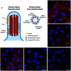
Figure 1. Primary cilia on hASC.
(a) The non-motile primary cilium axoneme is composed of 9 pairs of peripheral microtubule doublets arranged concentrically in 9+0 configuration, devoid of a central tubulin pair. (b–f) Primary cilia appear as a dense cluster of acetylated tubulin protruding from the apical surface of hASC in confluent culture. (b) Few cilia are observed 24 hours after culture in complete growth expansion media (CGM) in subconfluent culture. They are observed at day 3 (c) undifferentiated in CGM, (d) differentiated in osteogenic differentiation medium (ODM) and at day 12 in (e) undifferentiated in CGM and (f) differentiated in ODM. Cilia are identified with anti-acetylated tubulin (green) or in combination with CEP164 (red), actin identified with phalloidin (red) and nuclei are identified with DAPI staining (blue). Scale bar = 25 µm.
Figure 2
Figure 2. hASC expression of polycystin-1 (PC1), polycystin-2 (PC2) and intraflagellar transport protein-88 (IFT88).
(a) PC1 and (b) PC2 are observed as a diffuse signal on the cell body and at the base of the cilium. (c) IFT-88 is localized at the base and tip of the primary cilium. Cilia are identified with anti-acetylated tubulin (green), PC1, PC2 and IFT88 (red), and nuclei are identified with DAPI staining (blue). Scale Bar = 25 µm.
Figure 3
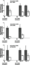
Figure 3. PC1, PC2 and IFT88 gene expression up to 15 days in ODM culture.
siRNA knockdown applied at days 0 and 7, confers at least a 50% reduction in mRNA expression of each cilia-associate protein (n = 3, Error bars = SEM).
Figure 4
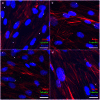
Figure 4. The effects of PC1, PC2 and IFT88 siRNA knockdown on hASC primary cilia.
hASC were cultured in expansion media (CGM) for 72 hours. Cilia expression on hASC: (a) Non-targeting scramble siRNA baseline cilia expression on approximately 50% of the hASC population (b) PC1 and (c) PC2 knockdown conditions express more cilia observed in a curved conformation and a minimal decrease in cilia population frequency, (d) IFT88 knockdown confers a reduction in cilia frequency on the hASC population. Cilia are identified with anti-acetylated tubulin (green), actin identified with phalloidin 594 (red) and nuclei are identified with DAPI staining (blue). Scale Bar = 25 µm.
Figure 5
Figure 5. The effect of knockdown on hASC proliferation in expansion media (CGM).
DNA was collected at days 0, 3, 7 and 10 to measure proliferative activity with siRNA knockdown of PC1, PC2 and IFT88 in as compared to the non-targeting scramble control siRNA. Knockdown of IFT88 significantly increases hASC proliferation as compared to control (n = 3, error bars = SEM, Student's t-test comparing each treatment to control, * p<0.05, ** p<0.01).
Figure 6
Figure 6. Osteogenic gene marker expression in hASC.
IFT88 knockdown confers a reduction in (a) Runx2 gene expression after 3 day and (c) BMP-2 gene expression after 10 day culture in osteogenic differentiation medium (ODM). (b) PC1, PC2 and IFT88 knockdown confer a reduction in alkaline phosphatase (ALP) gene expression after 10 days of culture in ODM. (d) PC1 knockdown confers downregulation of osteocalcin after 15 days of culture in osteogenic differentiation. Knockdown treatments applied at day 0 and day 7 of differentiation, (n = 3, error bars = SEM, Student's t-test comparing each treatment to control, * p<0.05, ** p<0.01).
Figure 7
Figure 7. Calcium accretion is affected by cilia-associated protein knockdown.
(a) Alizarin Red staining indicating degree of mineralization after 14 days ODM with knockdown. (b) Quantified calcium accretion normalized to DNA content. PC1 and PC2 knockdown leads to diminished calcium accretion, (n = 3, error bars = SEM, Student's t-test comparing each treatment to control, * p<0.05).
Figure 8
Figure 8. Endogenous alkaline phosphatase (ALP) activity of hASC after 14 days of culture.
hASC were cultured in osteogenic differentiation medium (ODM) and transfected with non-targeting scramble, PC1, PC2 and IFT88 siRNA. ODM and complete growth medium (CGM) hASC without knockdown (no KD) were also measured for comparison of baseline activity. Alkaline phosphatase activity is suppressed with cilia-associated protein knockdown. Knockdown of PC1 (*** p<0.01), PC2 (* p<0.05) and IFT88 (* p<0.05) all led to a significant decrease in ALP enzymatic activity compared to the non-targeting scramble siRNA knockdown control (n = 3, error bars = SEM, One-way repeated measures ANOVA, with Newman-Keuls post-hoc test).

References

    1. Singla V (2006) The Primary Cilium as the Cell's Antenna: Signaling at a Sensory Organelle. Science 313: 629–633 doi:10.1126/science.1124534. - DOI - PubMed
    1. Pazour GJ, Witman GB (2003) The vertebrate primary cilium is a sensory organelle. Current Opinion in Cell Biology 15: 105–110 doi:10.1016/S0955-0674(02)00012-1. - DOI - PubMed
    1. Santos N, Reiter JF (2008) Building it up and taking it down: The regulation of vertebrate ciliogenesis. Dev Dyn 237: 1972–1981 doi:10.1002/dvdy.21540. - DOI - PMC - PubMed
    1. Eggenschwiler JT, Anderson KV (2007) Cilia and Developmental Signaling. Annu Rev Cell Dev Biol 23: 345–373 doi:10.1146/annurev.cellbio.23.090506.123249. - DOI - PMC - PubMed
    1. Abou Alaiwi WA, Lo ST, Nauli SM (2009) Primary Cilia: Highly Sophisticated Biological Sensors. Sensors 9: 7003–7020 doi:10.3390/s90907003. - DOI - PMC - PubMed

Publication types

MeSH terms

Substances