. 2013 May 17;8(5):e62823.
doi: 10.1371/journal.pone.0062823. Print 2013.

Simvastatin inhibits renal cancer cell growth and metastasis via AKT/mTOR, ERK and JAK2/STAT3 pathway

Affiliations

Simvastatin inhibits renal cancer cell growth and metastasis via AKT/mTOR, ERK and JAK2/STAT3 pathway

Zhiqing Fang et al. PLoS One. .

Abstract

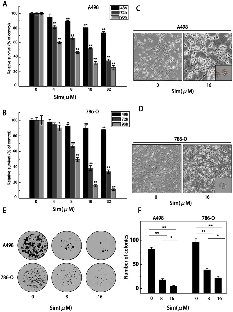
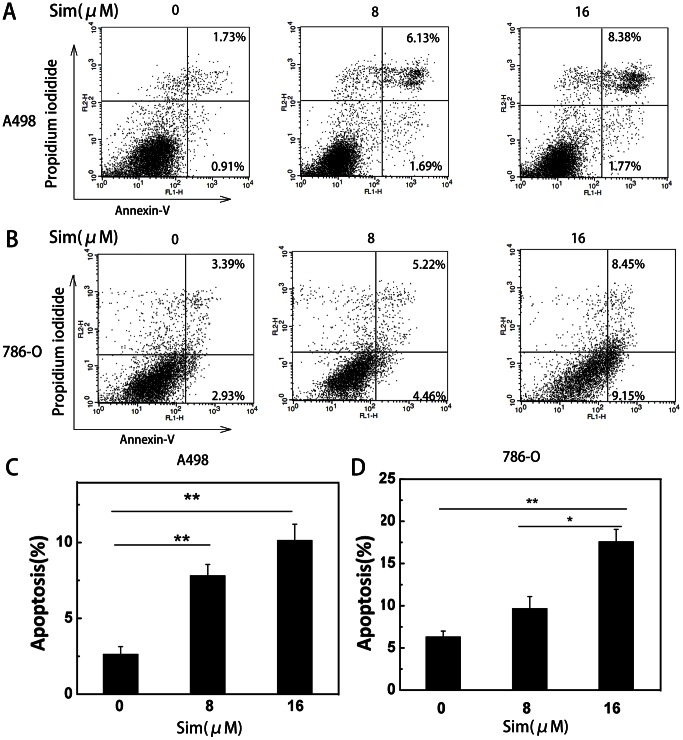
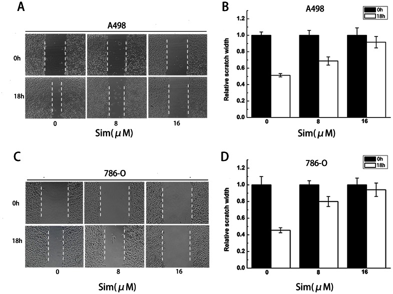
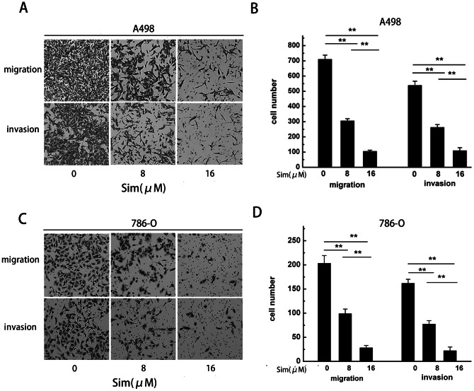
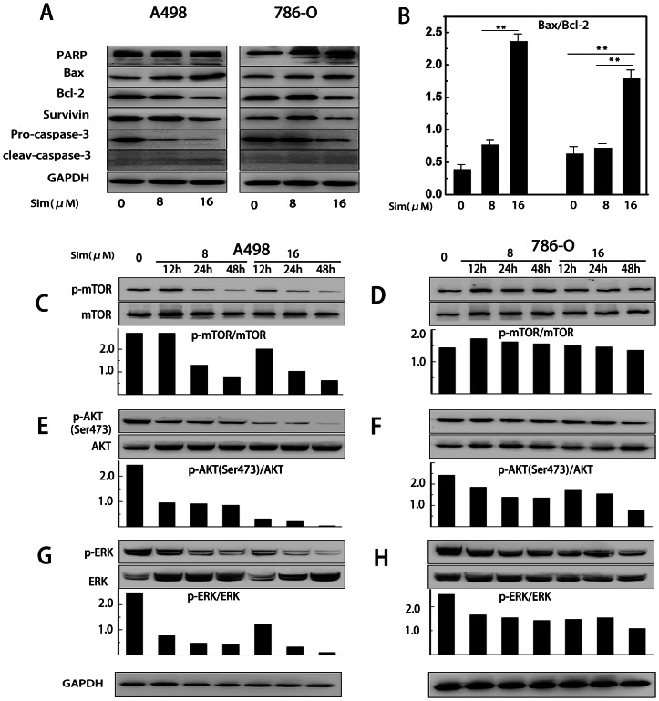
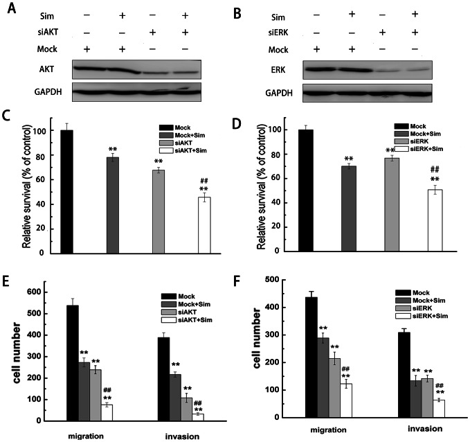
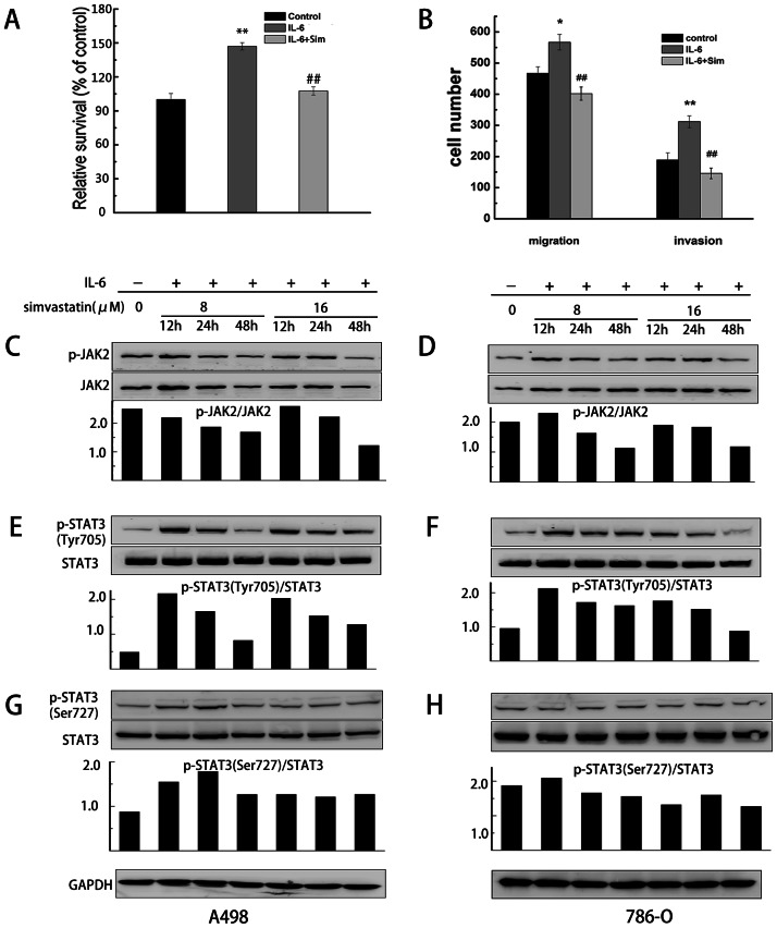
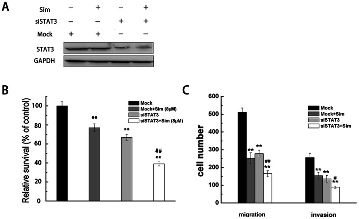
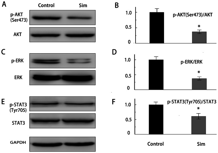
Renal cell carcinoma (RCC) is the most lethal type of genitourinary cancer due to its occult onset and resistance to chemotherapy and radiation. Recently, accumulating evidence has suggested stains, inhibitors of 3-hydroxy-3-methyl glutaryl coenzyme A (HMG-CoA) reductase, were associated with the risk reduction of cancer. In the present study, we aimed to investigate the potential effects of simvastatin on RCC cells and the underlying mechanisms by which simvastatin exerted its actions. With cell viability, colony formation, and flow cytometric apoptosis assays, we found that simvastatin potently suppressed cell growth of A498 and 786-O cells in a time- and dose- dependent manner. Consistently, the xenograft model performed in nude mice exhibited reduced tumor growth with simvastatin treatment. In addition, the inhibitory effects of simvastatin on migration and invasion were also observed in vitro. Mechanically, we presented that simvastatin could suppress the proliferation and motility of RCC cells via inhibiting the phosphorylation of AKT, mTOR, and ERK in a time- and dose- dependent manner. Further investigation of the underlying mechanism revealed simvastatin could exert the anti-tumor effects by suppressing IL-6-induced phosphorylation of JAK2 and STAT3. In conclusion, these findings suggested that simvastatin-induced apoptosis and its anti-metastasis activity in RCC cells were accompanied by inhibition of AKT/mTOR, ERK, and JAK2/STAT3 pathways, which imply that simvastatin may be a potential therapeutic agent for the treatment of RCC patients.


Conflict of interest statement

Competing Interests: The authors have declared that no competing interests exist.

Figures

Figure 1
Figure 1. Simvastatin suppressed cell viability of A498 and 786-O cells.
The effect of simvastatin on cell viability was measured by MTT assay. (A) A498 cells and (B) 786-O cells were treated with simvastatin for 48, 72 and 96 h. Simvastatin significantly inhibited cell viability of both cell lines in a dose-and time-dependent manner. Morphological changes of A498 and 786-O cells induced by simvastatin were displayed. Images of (C) A498 and (D) 786-O cells before and after addition of simvastatin (16 µm) were taken at 48 h. After treated with simvastatin for 48 h, apoptotic bodies in (C) A498 and (D) 786-O cells can be observed by optical microscope. (E) Inhibitory effect of simvastatin on colony formation. (F) The colony number was counted under microscope and colony is defined to consist of at least 50 cells. The results represent as mean ± SD of three independent experiments and the corresponding standard error. *P<0.05; **P<0.01.
Figure 2
Figure 2. Simvastatin induced dose-dependent apoptosis in A498 and 786-O cells.
(A) A498 and (B) 786-O cells were treated with simvastatin (0, 8 and 16 µM) for 48 hours and stained with FITC-annexinV and PI. The percentage of surviving cells was shown in the lower left quadrant; the percentage of early stage of apoptosis and late stage of apoptosis cells were shown in the lower right and upper right quadrants, respectively. (C, D) The quantification of apoptosis induced by simvastatin was calculated. Data is presented as mean ± SD of three independent experiments. * P<0.05, ** P<0.01.
Figure 3
Figure 3. The scratch assay showed the effect of simvastatin on cell migration.
(A) Scratch assay of A498 cells treated with 0, 8 and 16 µm of simvastatin. (B) The migration inhibition was transformed to the percentage of the initial distance between the two edges. The simvastatin-treated A498 cells showed a lower rate of wound closure than the control cells. (C) Scratch assay of 786-O cells treated with 0, 8 and 16 µm of simvastatin. (D) The simvastatin-treated 786-O cells showed a lower rate of wound closure than the control cells.
Figure 4
Figure 4. Cell migration and invasion ability were inhibited by increasing concentrations of simvastatin.
(A) Treatment with 0, 8 and 16 µm of simvastatin showed inhibited migration and invasion of A498 cells. (B) The number of A498 cells that successfully migrated and invaded was counted. (C) Migration and invasion of 786-O cells was inhibited after treatment with different concentrations of simvastatin. (D) The decreased number of 786-O cells indicated the great inhibitory effect of simvastatin on cell mobility. Data is presented as mean ± SD of three independent experiments. * P<0.05, ** P<0.01.
Figure 5
Figure 5. Simvastatin inhibited the growth and induced cell apoptosis in RCC tumor xenografts.
Images of the excised tumors (A) and the nude mice (C) were taken from the control and treatment group. The arrows point to the xenografts. (B) Graphs representing the average tumor volumes of A498 xenografts treated with or without simvastatin. (D) Body weight curve of nude mice bearing A498 tumors treated with simvastatin. (E) Representative TUNEL staining (red fluoresence) of A498 renal cancer xenografts. (F) Bar graph showing quantification of the TUNEL positive A498 cells in tumor xenogafts. Data are presented as mean ± SD, *p<0.05.
Figure 6
Figure 6. Effects of simvastatin on the protein levels of Bcl-2, Bax, caspase 3, PARP, mTOR, AKT, and ERK in A498 and 786-O cells.
(A) A498 and 786-O cells were assayed for Bcl-2, Bax, full length caspase 3 and cleaved caspase 3, full length PARP and cleaved PARP by western blotting analysis with GAPDH as a control. (B) Bax/Bcl-2 ratios of A498 and 786-O cells. The densitometry value of each band was determined with ImageJ. Data was presented as mean ± SD of three independent experiments. * P<0.05, ** P<0.01. (C–H) The levels of mTOR, AKT, ERK and their phosphorylated forms were analyzed by western blotting. Quantitation of the p-mTOR/mTOR, p-AKT/AKT and p-ERK/ERK ratio was determined by densitometry analysis.
Figure 7
Figure 7. Simvastatin depressed proliferation and metastasis of A498 cells that were transfected with AKT or ERK siRNA compared with the control cells (Mock).
(A, B) A498 cells were transfected and treated with simvastatin (8 µM), and the levels of AKT and ERK were analyzed by western blotting with GAPDH as a control. After transfected with AKT or ERK siRNA, A498 cells were incubated in the absence or presence of simvastatin (8 µM) for 48 h. The cell viability was measured by MTT assay (C, D), and cell migration and invasion was measured by transwell assay (E, F). * p<0.05 or ** p<0.01, compared with the untreated cells (Mock). # p<0.05 or ## p<0.01, compared with the cells transfected with AKT or ERK siRNA.
Figure 8
Figure 8. Simvastatin inhibited IL-6 induced proliferation, migration and invasion of A498 cells via inhibition of JAK2/STAT3 pathway.
(A, B) A498 cells were treated with IL-6 (10 ng/ml) for 48 h, and cell vitality and motility was estimated using the MTT and transwell assay respectively. Each bar represents the mean±SD of three independent experiments. *p<0.05 or ** p<0.01, compared with cells treated without IL-6. (C–H) Simvastatin inhibited the phosphorylation of Jak2, STAT3 (Tyr705) and STAT3 (Ser727) in renal cancer cells in a dose- and time-dependent manner. Quantitative analysis of the p-JAK2/JAK2, p-STAT3 (Tyr705)/STAT3 and p-STAT3 (Ser727)/STAT3 ratio was displayed in each lower panel.
Figure 9
Figure 9. Simvastatin suppressed proliferation and metastasis of A498 cells that were transfected with STAT3 siRNA.
(A) A498 cells were transfected with STAT3 siRNA and treated with simvastatin (8 µM), and the levels of STAT3 was analyzed by western blotting with GAPDH as a control. (B, C) After transfected with STAT3 siRNA, A498 cells were incubated in the absence or presence of simvastatin (8 µM) for 48 h. The cell viability was measured by MTT assay, (B), and cell migration and invasion was measured by transwell assay (C). * p<0.05 or ** p<0.01, compared with the untreated cells (Mock). # p<0.05 or ## p<0.01, compared with the cells transfected with STAT3 siRNA.
Figure 10
Figure 10. Anti-tumor effects of simvastatin on A498 tumor xenograft were associated with the inhibition of AKT, ERK and STAT3 activity.
Western blotting analysis for the phosphorylated AKT, ERK and STAT3 levels in tumor xenograft collected from nude mice treated with or without simvastatin. Quantitation of p-AKT/AKT, p-ERK/ERK and p-STAT3/STAT3 ratio was determined by densitometry analysis. *P<0.05 compared with control.

References

    1. Bex A, Jonasch E, Kirkali Z, Mejean A, Mulders P, et al. (2010) Integrating surgery with targeted therapies for renal cell carcinoma: current evidence and ongoing trials. Eur Urol 58: 819–828. - PubMed
    1. Basso M, Cassano A, Barone C (2010) A survey of therapy for advanced renal cell carcinoma. Urol Oncol 28: 121–133. - PubMed
    1. Lam JS, Leppert JT, Belldegrun AS, Figlin RA (2005) Novel approaches in the therapy of metastatic renal cell carcinoma. World J Urol 23: 202–212. - PubMed
    1. Linehan WM, Zbar B (2004) Focus on kidney cancer. Cancer Cell 6: 223–228. - PubMed
    1. Motzer RJ, Russo P (2000) Systemic therapy for renal cell carcinoma. J Urol 163: 408–417. - PubMed

Publication types

MeSH terms

LinkOut - more resources