. 2013 May 15;8(5):e63364.
doi: 10.1371/journal.pone.0063364. Print 2013.

The identification of gut neuroendocrine tumor disease by multiple synchronous transcript analysis in blood

Affiliations

The identification of gut neuroendocrine tumor disease by multiple synchronous transcript analysis in blood

Irvin M Modlin et al. PLoS One. .

Abstract

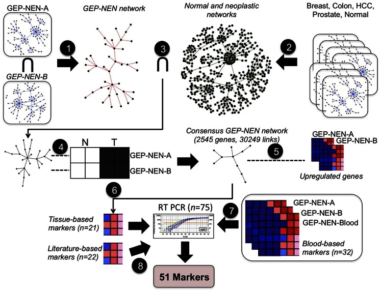
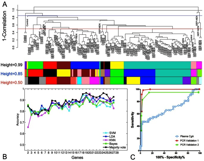
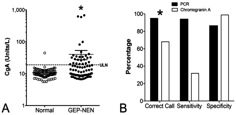
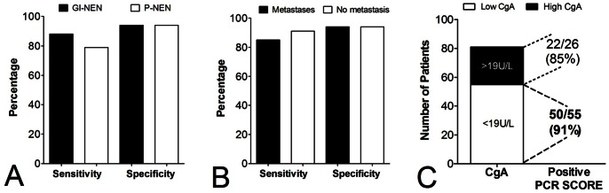
Gastroenteropancreatic (GEP) neuroendocrine neoplasms (NENs) are increasing in both incidence and prevalence. A delay in correct diagnosis is common for these lesions. This reflects the absence of specific blood biomarkers to detect NENs. Measurement of the neuroendocrine secretory peptide Chromogranin A (CgA) is used, but is a single value, is non-specific and assay data are highly variable. To facilitate tumor detection, we developed a multi-transcript molecular signature for PCR-based blood analysis. NEN transcripts were identified by computational analysis of 3 microarray datasets: NEN tissue (n = 15), NEN peripheral blood (n = 7), and adenocarcinoma (n = 363 tumors). The candidate gene signature was examined in 130 blood samples (NENs: n = 63) and validated in two independent sets (Set 1 [n = 115, NENs: n = 72]; Set 2 [n = 120, NENs: n = 58]). Comparison with CgA (ELISA) was undertaken in 176 samples (NENs: n = 81). 51 significantly elevated transcript markers were identified. Gene-based classifiers detected NENs in independent sets with high sensitivity (85-98%), specificity (93-97%), PPV (95-96%) and NPV (87-98%). The AUC for the NEN gene-based classifiers was 0.95-0.98 compared to 0.64 for CgA (Z-statistic 6.97-11.42, p<0.0001). Overall, the gene-based classifier was significantly (χ(2) = 12.3, p<0.0005) more accurate than CgA. In a sub-analysis, pancreatic NENs and gastrointestinal NENs could be identified with similar efficacy (79-88% sensitivity, 94% specificity), as could metastases (85%). In patients with low CgA, 91% exhibited elevated transcript markers. A panel of 51 marker genes differentiates NENs from controls with a high PPV and NPV (>90%), identifies pancreatic and gastrointestinal NENs with similar efficacy, and confirms GEP-NENs when CgA levels are low. The panel is significantly more accurate than the CgA assay. This reflects its utility to identify multiple diverse biological components of NENs. Application of this sensitive and specific PCR-based blood test to NENs will allow accurate detection of disease, and potentially define disease progress enabling monitoring of treatment efficacy.


Conflict of interest statement

Competing Interests: We received funding from a commercial source: Clifton Life Sciences. This does not alter our adherence to all the PLOS ONE policies on sharing data and materials.

Figures

Figure 1
Figure 1. Computational pipeline used to derive a set of 51 markers that identify GEP-NEN disease.
Step 1: Gene co-expression networks inferred from GEP-NEN-A and GEP-NEN-B datasets are intersected, producing the GEP-NEN network. Step 2: Co-expression networks from neoplastic and normal tissue microarray datasets are combined to produce the normal and neoplastic networks. Step 3: Links present in normal and neoplastic networks are subtracted from the GEP-NEN network. Step 4: Concordantly regulated genes in GEP-NEN-A and GEP-NEN-B networks are retained; other genes are eliminated from the GEP-NEN network, producing the Consensus GEP-NEN network. Step 5: Upregulated genes in both the GEP-NEN-A and GEP-NEN-B dataset are mapped to the Consensus GEP-NEN network. Step 6: Topological filtering, expression profiling, and literature-curation of putative tissue-based markers, yielding 21 putative genes further examined by RT-PCR. Step 7: Identification of mutually up-regulated genes in GEP-NEN blood transcriptome and GEP-NEN-A and GEP-NEN-B datasets, yielding 32 putative genes further examined by RT-PCR. Step 8: Literature-curation and cancer mutation database search, yielding a panel of 22 putative marker genes for further RT-PCR analysis.
Figure 2
Figure 2. GEP-NEN gene co-expression network.
A) Visualization of the GEP-NEN gene co-expression network (2545 genes, 30249 edges). Each node represents a gene, while a link represents a GEP-NEN-specific co-expression. Nodes that localized to the same network community are marked in the same color. The community structure of the GEP-NEN network is further visualized in the 3 dimensional inset, whereby each node represents a community while edges are drawn between communities that contain co-expressed genes. Larger nodes indicate bigger gene communities. B) Heatmap visualizing enrichment for over-represented Gene Ontology (GO) Biological Process (BP) terms assigned to the 10 largest clusters (>20 genes). Heatmap colors represent the significance of the enrichment.
Figure 3
Figure 3. Utility of the 51 marker gene signature for identification of GEP-NEN disease.
A) Unsupervised hierarchical clustering of the 130 samples in the training set (n = 67 controls, n = 63 GEP-NENs). Tree was generated with an average agglomeration method and 1-(sample correlation) was used as a measure of dissimilarity. Unique colors under the dendrogram represent sample cluster assignments, computed by cutting the hierarchical tree at height = 0.99 (black line), 0.85 (blue line), or 0.50 (red line) using a dynamic tree cutting approach . B) Prediction accuracy of each classifier using sequential addition of 27 significantly up-regulated genes (p<0.05) in the GEP-NEN samples obtained using results of the 10-fold cross validation. C) Receiver operating characteristic (ROC) curves for “majority vote” classifier applied to validation sets 1 (AUC = 0.98, p<0.0001) and 2 (AUC = 0.95, p<0.0001) compared to ROC curve for utility of the plasma CgA values (AUC = 0.64, p<0.002). Direct comparisons of AUCs between set 1 or set 2 and CgA identified estimated Z-scores of 10.57 and 11.42 respectively, confirming the significant differences between the two detection systems (calculations detailed in Supplementary Methods ).
Figure 4
Figure 4. Comparison of the 51 marker gene signature with Chromogranin A (CgA) for detecting GEP-NENs.
A) CgA levels were significantly elevated in the GEP-NEN group (n = 176; *p<0.002) but an overlap with normal values was identified. B) Comparison of the PCR-based approach with CgA protein measurement identified that call rates were significantly higher for the PCR-based test (*p<0.0005, χ2 = 12.3). The PCR blood test was significantly more accurate than measurement of CgA levels to detect GEP-NENs. ULN = upper limit of normal (19U/L – DAKO).
Figure 5
Figure 5. Utility of the 51 marker gene signature for detecting P-NENs, metastases and in patients with low Chromogranin A (CgA).
A) The sensitivity and specificity of the test to detect GI-NENs (90%, 94%) and P-NENs (80%, 94%) was similar. B) The PCR-based approach could detect patients with no metastases as well as patients with metastases. C) The PCR-based test could accurately identify GEP-NENs even when plasma CgA were low (<19U/L). Overall, the PCR blood test was significantly more accurate than measurement of CgA levels to detect GEP-NENs (*p<10−13, χ2>50).

References

    1. Modlin IM, Oberg K, Chung DC, Jensen RT, de Herder WW, et al. (2008) Gastroenteropancreatic neuroendocrine tumours. Lancet Oncol 9: 61–72. - PubMed
    1. Yao JC, Hassan M, Phan A, Dagohoy C, Leary C, et al. (2008) One hundred years after “carcinoid”: epidemiology of and prognostic factors for neuroendocrine tumors in 35,825 cases in the United States. J Clin Oncol 26: 3063–3072. - PubMed
    1. Kulke MH, Siu LL, Tepper JE, Fisher G, Jaffe D, et al. (2011) Future directions in the treatment of neuroendocrine tumors: consensus report of the National Cancer Institute Neuroendocrine Tumor clinical trials planning meeting. J Clin Oncol 29: 934–943. - PMC - PubMed
    1. Yao JC, Pavel M, Phan AT, Kulke MH, Hoosen S, et al. (2011) Chromogranin A and neuron-specific enolase as prognostic markers in patients with advanced pNET treated with everolimus. J Clin Endocrinol Metab 96: 3741–3749. Epub 2011 Oct 3712. - PubMed
    1. AACC (2010) Chromogranin A. Lab Tests Online: American Association for Clinical Chemistry. pp. Details regarding Chromogranin A as a test.

Publication types