. 2013 May 14;8(5):e63843.
doi: 10.1371/journal.pone.0063843. Print 2013.

Septins are important for cell polarity, septation and asexual spore formation in Neurospora crassa and show different patterns of localisation at germ tube tips

Affiliations

Septins are important for cell polarity, septation and asexual spore formation in Neurospora crassa and show different patterns of localisation at germ tube tips

Adokiye Berepiki et al. PLoS One. .

Abstract

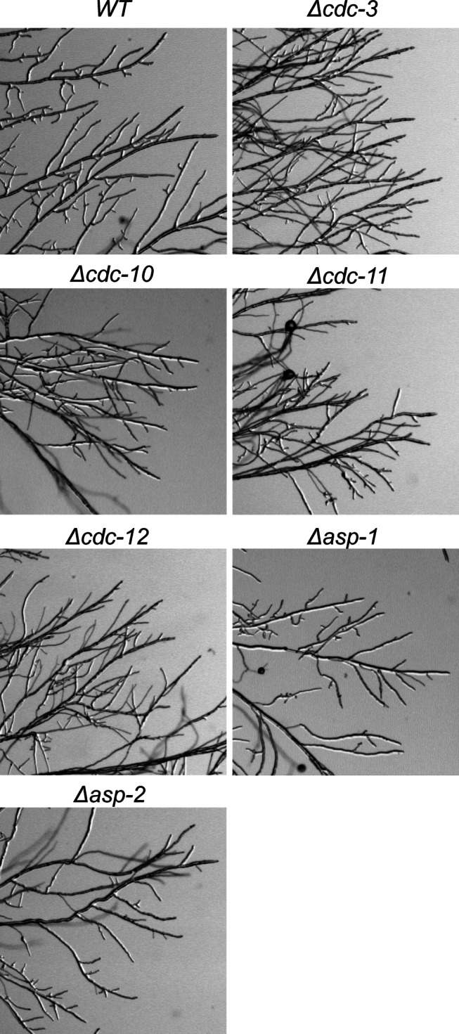
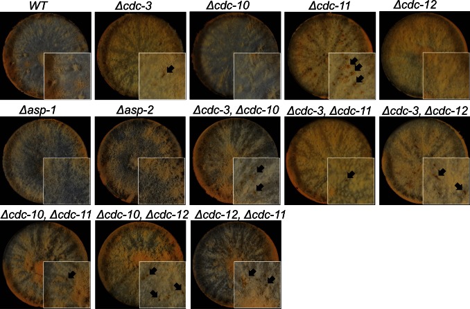
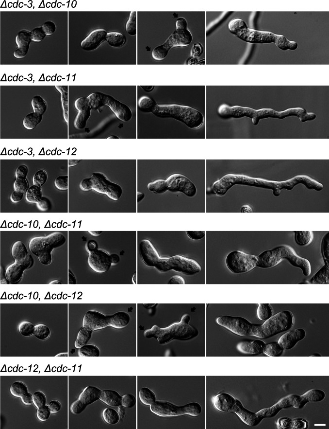
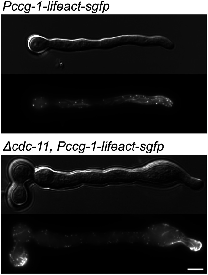
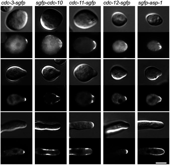
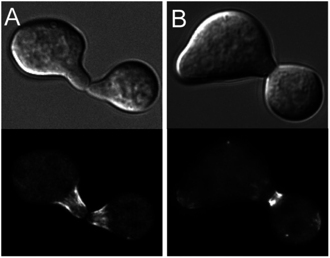
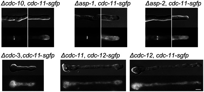
Septins are GTP-binding cytoskeletal proteins that contribute to cell polarity, vesicle trafficking, cytokinesis and cell morphogenesis. Here we have characterised the six septins encoded by the genome of the model filamentous fungus Neurospora crassa. Analysis of septin null mutants demonstrated that septins limit the sites of emergence of germ tubes and are important for septation and conidiation in N. crassa. Septins constituted a range of different higher-order structures in N. crassa - rings, loops, fibres, bands, and caps - which can co-exist within the same cell. They showed different patterns of localisation at germ tube tips, with GFP-CDC-10 and CDC-11-GFP forming a subapical collar with lower signal intensity at the tip apex, CDC-3-GFP and CDC-12-GFP organized as a cap at the tip apex and GFP-ASP-1 forming an extended subapical collar. Purification of the septin complex and mass spectrometry of isolated proteins revealed that the septin complex consists predominantly of CDC-3, CDC-10, CDC-11 and CDC-12. Immunoprecipitation of the putative septin ASP-1 revealed that this protein interacts with the core septin complex.


Conflict of interest statement

Competing Interests: The authors have declared that no competing interests exist.

Figures

Figure 1
Figure 1. Septin deletion strains have altered morphologies.
Wild-type and single septin deletion strains were incubated in liquid VMM for up to 5 h then imaged by DIC microscopy to obtain representative images of germination and development. Septin deletion strains, with the exception of Δasp-1 and Δasp-2, fail to properly separate conidia and often appeared swollen and misshapen. Black arrows denote multiple germ tube emergence. White arrows show multiple CAT emergence. Scale bar, 5 µm.
Figure 2
Figure 2. Septin deletion strains display an increase in germ tube emergence and unseparated conidia and a reduction in septation compared to wild-type.
(A) The number of unseparated cells in was counted before incubation, the number of cells with more than two germ tubes was counted following 3 h of incubation in liquid VMM and the number of septate cells was determined with DIC microscopy after 5 h of incubation in liquid VMM (n = 300). There was a significant increase (p = <0.05) in the percentage of unseparated conidia and cells with multiple germ tubes compared to wild-type for all strains with the exception of Δcdc10, Δasp-1 and Δasp-2. Similarly, there was a significant decrease (p = <0.05) in the percentage of septate cells for all strains compared to the wild-type except for Δasp-1 and Δasp-2. (B) Strains were incubated in liquid VMM for 3 h then quantified for germination and cell fusion (n = 300). A black dot above bars indicate a significance difference (p = <0.05) compared to the wild-type control. The germination rates of Δcdc-10 and Δcdc-11 were slightly reduced compared to the wild-type.
Figure 3
Figure 3. CAT-mediated cell fusion is affected in septin null mutants.
Wild-type and single septin deletion strains were incubated in liquid VMM for up to 5 h then imaged by DIC microscopy to obtain representative images of cell fusion. In the wild-type each conidium most commonly formed a single CAT. In septin deletion strains, often several CATs are formed during cell fusion. Black arrows denote individual CATs prior to fusion. Scale bar, 5 µm.
Figure 4
Figure 4. Branching is more prevalent at the colony edge in septin null mutants.
Wild-type and septin deletion strains were inoculated onto solid VMM, incubated for 18 d at 35°C then imaged using a stereomicroscope. Septin deletion strains, with the exception of Δasp1-1 and Δasp-2, appeared to form more branches at the colony edge.
Figure 5
Figure 5. Colony morphology is altered in septin deletion strains.
Wild-type and septin deletion strains were inoculated onto solid VMM, incubated for 3 d at 24°C then photographed. Septin deletion strains, with the exception of Δasp1-1 and Δasp-2, formed denser colonies, did not produce as many aerial hyphae as wild-type and occasionally regions of cell lysis were visible (black arrows).
Figure 6
Figure 6. Septin double deletion strains have altered morphologies.
Wild-type and septin deletion strains were incubated in liquid VMM for up to 5 h then imaged by DIC microscopy to obtain representative images of germination and development. Septin double deletion strains were swollen and misshapen and failed to properly separate conidia. Black arrows denote multiple germ tube emergence. Scale bar, 5 µm.
Figure 7
Figure 7. F-actin structures are unaffected in the absence of cdc-11 .
Lifeact-GFP-expressing wild-type and Δcdc-11 strains were incubated in liquid VMM for 3 h then imaged by widefield fluorescence microscopy. F-actin structures appeared as normal although due to the polarity defects of the cdc-11 null mutant, and actin accumulated at the tip and in the spore body. Scale bar, 5 µm.
Figure 8
Figure 8. Septins show different patterns of localisation at germ tube tips.
Septin-GFP strains were incubated in liquid VMM for 3 h then imaged with DIC and widefield fluorescence microscopy to obtain representative images of germination and development. At the hyphal tip septins localised as a cap (CDC-3-GFP and CDC-12-GFP), an extended cap (GFP-ASP-1) or as a bar-like structures (GFP-CDC-10 and CDC-11-GFP). Scale bar, 5 µm.
Figure 9
Figure 9. CDC-11 localises to the tips of conidial anastomosis tubes (CATs) and becomes concentrated around the fusion pore.
Cells expressing CDC-11-GFP were incubated in liquid VMM for 3 h then imaged with DIC and widefield fluorescence microscopy. (A) CDC-11-GFP localised as bar-like structures within the tips of CATs before and after making contact with each other. (B) After cell fusion, CDC-11 became concentrated around the fusion pore. Scale bar, 5 µm.
Figure 10
Figure 10. Septins form diverse HO structures.
Cells expressing GFP-tagged septins were incubated in liquid VMM for 3 h then imaged with DIC and widefield fluorescence microscopy to obtain representative images of HO structures and septin ring formation. Septin rings formed by CDC-3-GFP (A), GFP-CDC-10 (B), CDC-11-GFP (C), CDC-12-GFP (D), and GFP-ASP-1(E). (F1, F2, F3) The CDC-11-GFP-labelled septin ring was formed by the accumulation of septin fibres. (G) Dense septin loops formed by CDC-11-GFP were associated with the cell cortex of an ungerminated conidium. (H, I) Septin fibres formed by GFP-CDC-10 (H) and CDC-11-GFP (I) were present in the cytoplasm. Scale bar, 5 µm.
Figure 11
Figure 11. Septins fibres are more abundant when GFP-CDC-10 is overexpressed.
Conidia from two GFP-CDC-10 strains were harvested then imaged with fluorescence and DIC microscopy to obtain representative images of septin fibres. When GFP-CDC-10 is expressed from the Pccg-1 promoter instead of its native promoter then septin fibres are more abundant. The higher fluorescence background in sgfp-cdc-10 was due to the lower expression of the native cdc10 promoter compared with the ccg-1 promoter. Scale bar, 5 µm.
Figure 12
Figure 12. Higher order septin structures fail to form in the absence of CDC-3, CDC-11, or CDC-12.
Septin deletion strains expressing CDC-11-GFP or CDC-12-GFP were incubated in liquid VMM for 5 h then imaged with DIC microscopy and widefield fluorescence to obtain representative images of septin ring formation in the absence of a core septin. Scale bar, 5 µm.
Figure 13
Figure 13. Identification of constituents of septin complexes.
SYPRO Ruby-stained SDS PAGE gel of septin complexes. Protein were purified from septin-V5-HAT expressing strains and identified by mass spectrometry. The positions of the molecular mass markers (M, in kDa) are shown at the left of the panel.

References

    1. Nishihama R, Onishi M, Pringle JR (2011) New insights into the phylogenetic distribution and evolutionary origins of the septins. Biol Chem 392: 681–687. - PMC - PubMed
    1. Pan F, Malmberg RL, Momany M (2007) Analysis of septins across kingdoms reveals orthology and new motifs. BMC Evol Biol 7: 103–109. - PMC - PubMed
    1. Hartwell LH (1971) Genetic control of the cell division cycle in yeast. IV. Genes controlling bud emergence and cytokinesis. Exp Cell Res 69: 265–276. - PubMed
    1. Spiliotis ET, Gladfelter AS (2012) Spatial guidance of cell asymmetry: septin GTPases show the way. Traffic 13: 195–203. - PMC - PubMed
    1. Sirajuddin M, Farkasovsky M, Hauer F, Kuhlmann D, Macara IG, et al. (2007) Structural insight into filament formation by mammalian septins. Nature 449: 311–315. - PubMed

Publication types