. 2013 May 17;8(5):e64232.
doi: 10.1371/journal.pone.0064232. Print 2013.

The minimal Bacillus subtilis nonhomologous end joining repair machinery

Affiliations

The minimal Bacillus subtilis nonhomologous end joining repair machinery

Miguel de Vega. PLoS One. .

Abstract

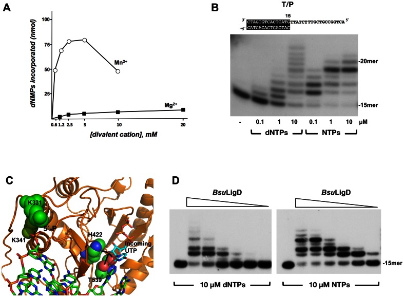
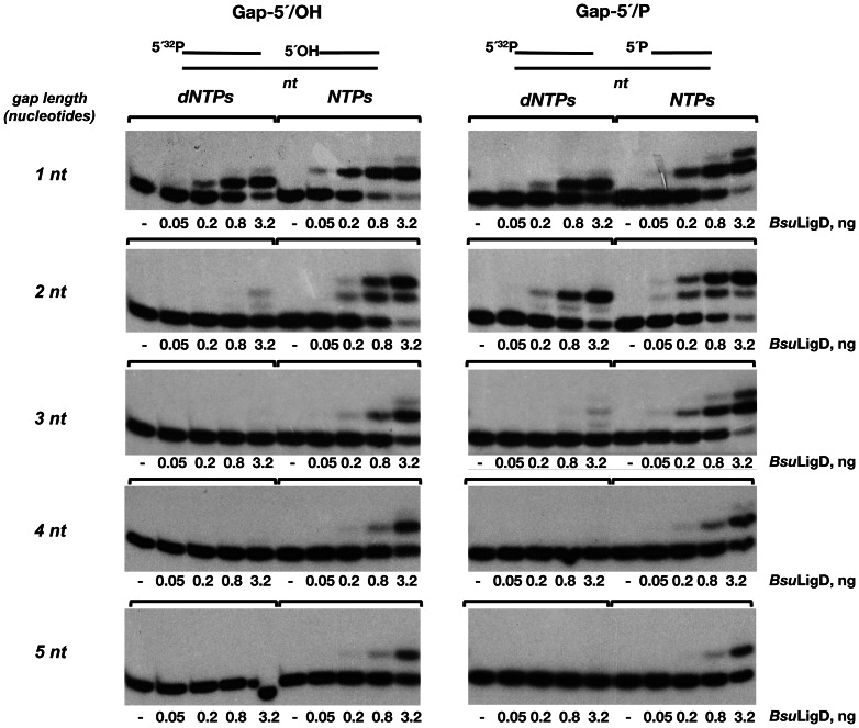
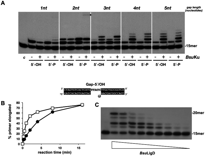
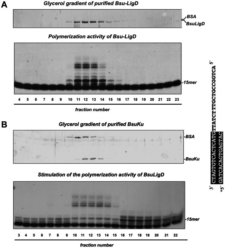
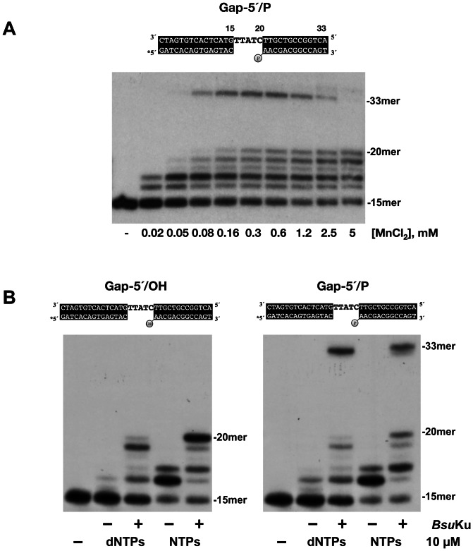
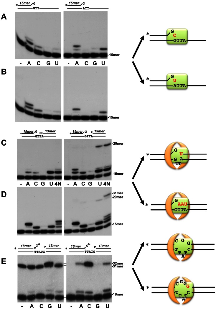
It is widely accepted that repair of double-strand breaks in bacteria that either sporulate or that undergo extended periods of stationary phase relies not only on homologous recombination but also on a minimal nonhomologous end joining (NHEJ) system consisting of a dedicated multifunctional ATP-dependent DNA Ligase D (LigD) and the DNA-end-binding protein Ku. Bacillus subtilis is one of the bacterial members with a NHEJ system that contributes to genome stability during the stationary phase and germination of spores, having been characterized exclusively in vivo. Here, the in vitro analysis of the functional properties of the purified B. subtilis LigD (BsuLigD) and Ku (BsuKu) proteins is presented. The results show that the essential biochemical signatures exhibited by BsuLigD agree with its proposed function in NHEJ: i) inherent polymerization activity showing preferential insertion of NMPs, ii) specific recognition of the phosphate group at the downstream 5' end, iii) intrinsic ligase activity, iv) ability to promote realignments of the template and primer strands during elongation of mispaired 3' ends, and v) it is recruited to DNA by BsuKu that stimulates the inherent polymerization and ligase activities of the enzyme allowing it to deal with and to hold different and unstable DNA realignments.


Conflict of interest statement

Competing Interests: The author has declared that no competing interests exist.

Figures

Figure 1
Figure 1. BsuLigD is endowed with a polymerization activity.
(A) Effect of Mg2+ and Mn2+ concentration on the polymerization activity of BsuLigD. The assay was performed as described in Materials and Methods, using as substrate 1.5 µg of activated calf thymus DNA, 400 ng of BsuLigD, 100 nM dNTPs and the indicated concentrations of MgCl2 or MnCl2. Polymerization activity was calculated as the amount of incorporated dNMP (nmol). (B) Polymerization activity of BsuLigD on a template/primer (T/P) DNA substrate. The molecule used as substrate in the analysis is depicted on top of the figure (see also Materials and Methods). Asterisk indicates the 5′32P-labeled end of the primer strand. Reactions were performed as described under Materials and Methods, using 1.5 nM of each substrate, 100 ng of BsuLigD, 5 mM MnCl2 and the indicated concentrations of nucleotides. After incubation for 20 min at 30°C, samples were analyzed by 8 M urea and 20% PAGE and autoradiography. (C) Ribbon representation of the structural model of BsuLigD polymerization domain (residues 320–611). Model was provided by the homology-modelling server Swiss-Model –, using as template the crystallographic structure of the MtuLigD preternary complex [PDB code 3PKY [42]]. BsuLigD residues K331 and K341, potentially involved in the specific recognition of the downstream 5′-P group are shown as lime and green space-filling spheres, respectively. The 2′-OH group of the incoming UTP is proposed to make hydrogen bonds (black lines) with BsuLigD residues H422 and T539, represented as space-filling spheres. Figure was made using PyMOL software (http://www.pymol.org). (D) BsuLigD shows a distributive polymerization pattern. The assay was carried out as described in Materials and Methods, using T/P substrate depicted on top of the part B, in the presence of 10 µM of the indicated type of nucleotides and decreasing amounts of BsuLigD (50, 25, 6, 2, 0.4 and 0.1 ng). After incubation for 20 min at 30°C, samples were analyzed by 8 M urea-20% PAGE and autoradiography.
Figure 2
Figure 2. Influence of the gap length and the 5′ group of the downstream strand in the gap-filling efficiency of Bsu LigD.
The assay was performed essentially as described in Materials and Methods. The indicated amounts of BsuLigD were incubated with 2.5 nM of the indicated gapped molecule in the presence of 100 µM of the specified nucleotide and 5 mM MnCl2. After incubation for 5 min at 30°C, samples were analyzed by 8 M urea-20% PAGE and autoradiography.
Figure 3
Figure 3. BsuKu interacts functionally with BsuLigD enhancing its gap-filling efficiency.
(A) Effect of BsuKu on the gap-filling efficiency of BsuLigD. The assay was performed as described in Materials and Methods, incubating 0.4 ng of BsuLigD with 2.5 nM of the indicated gapped molecule, 5 mM MnCl2 and 1 µM NTPs, in the absence (−) or presence (+) of 40 ng of BsuKu. The 5′ end of the downstream strand is specified (5′-OH, 5′-P). After incubation for 10 min at 30°C, samples were analyzed by 8 M urea and 20% PAGE and autoradiography. (B) BsuKu increases BsuLigD primer usage. The assay was performed as described in Materials and Methods, incubating 10 ng of BsuLigD with 1 nM of the depicted gapped molecule, 5 mM MnCl2 and 10 µM NTPs, in the absence (full circles) or presence (squares) of 40 ng of BsuKu. After incubation for the indicated times at 30 °C, samples were analyzed by 8 M urea and 20% PAGE and autoradiography and the unextended and elongated primer molecules quantified using a Molecular Dynamics PhosphorImager. Percentage of extended primers (elongated/elongated+unextended) was plotted against reaction time. (C) BsuKu does not prevent BsuLigD dissociation after each NMP insertion reaction. The assay was carried out as described in Materials and Methods, using the 5-nt gapped molecule depicted, in the presence of 40 ng of BsuKu, 10 µM NTPs and decreasing amounts of BsuLigD (2.5, 1.25, 0.6, 0.3, 0.15, 0.07, 0.04, 0.02 and 0.01 ng). After incubation for 20 min at 30°C, samples were analyzed by 8 M urea-20% PAGE and autoradiography.
Figure 4
Figure 4. Cosedimentation of polymerization activity with BsuLigD.
(A) Top panel shows a SDS-PAGE analysis followed by Coomassie Blue staining of gradient fractions 4–23 collected after sedimentation of the purified BsuLigD on a 15–30% glycerol gradient (see Materials and Methods). Bottom panel shows the polymerization products obtained after incubating for 20 min at 30°C 4 µl of each fraction with the template/primer structure depicted on the right (see Materials and Methods), in the presence of 5 mM MnCl2, 100 µM dNTPs and 50 ng of BsuKu. Asterisk indicates the 32P 5′-labeled end of the primer strand. (B) Stimulation of the BsuLigD polymerization activity cosediments with BsuKu. Top panel shows a SDS-PAGE analysis followed by Coomassie Blue staining of gradient fractions 3–22 collected after sedimentation of the purified BsuKu on a 15–30% glycerol gradient (see Materials and Methods). Bottom panel shows the polymerization products obtained after incubating for 20 min at 30°C 50 ng of BsuLigD with the template/primer structure depicted on (A) (see Materials and Methods), in the presence of 5 mM MnCl2, 100 µM dNTPs and 4 µl of each fraction.
Figure 5
Figure 5. Complete repair of a gapped DNA substrate by the BsuLigD/BsuKu complex.
(A) Optimal metal requirement to couple polymerization and ligation activities of BsuLigD/BsuKu complex. The gap-filling assay was performed as described in Materials and Methods by incubating 50 ng of BsuLigD and BsuKu with 1.5 nM of the gapped DNA molecule depicted on top, in the presence of the indicated concentrations of MnCl2 and 1 µM NTPs. After incubation for 10 min at 30 °C, the reactions were stopped by adding EDTA up to 10 mM. Samples were analyzed by 8 M urea, 20% PAGE and autoradiography. Asterisk indicates the 5′32P-labeled end of the primer strand. (B) Polymerization and ligase activities of BsuLigD allow complete repair of a gapped molecule. The different molecules used in the analysis are depicted on top of each panel. The assay was performed as described in Materials and Methods by incubating the indicated gapped substrates depicted on top of the figure with 10 ng of BsuLigD, 10 µM of either dNTPs or NTPs, 0.6 mM MnCl2, in absence (−) or presence (+) of 40 ng of BsuKu. After incubation for 20 min at 30°C, the primer-extended products corresponding either to the filling-in reaction or to the complete repair reaction (filling-in + ligation) were analyzed by 8 M urea-20% PAGE and autoradiography.
Figure 6
Figure 6. Activity of BsuLigD/Ku complex on molecules bearing a mispaired 3′ end.
(A) and (B) Elongation of dG:dG and dG:dA mispairs present in a template/primer structure. The assay was performed essentially as described in Materials and Methods in the presence of 1 µM of the indicated nucleotide, 0.6 mM MnCl2, 1.5 nM of the depicted substrate and 50 ng of BsuLigD, in the absence (A) or presence (B) of 50 ng of BsuKu. After incubation for 20 min at 30°C the elongation and ligation products were analyzed by 8 M urea-20% PAGE and autoradiography. Position of the unextended primer is indicated on the right. Asterisks indicate the 32P 5′-labeled end of the primer strand. (C) and (D) Activity on a mispaired 3′ end in the 3 nucleotides gapped molecule. The assay was performed essentially as described in Materials and Methods in the presence of 1 µM of the indicated nucleotide (4N: the four nucleotides), 0.6 mM MnCl2, 1.5 nM of the depicted gapped substrate and 50 ng of BsuLigD, in the absence (C) or presence (D) of 50 ng of BsuKu. After incubation for 20 min at 30°C the elongation and ligation products were analyzed by 8 M urea-20% PAGE and autoradiography. Position of the unextended primer and ligation products is indicated on the right. (E) Activity on a 3 nucleotides mismatched 3′ terminus present in 5 nucleotides gapped molecules. The assay was performed essentially as described in Materials and Methods in the presence of 1 µM of the indicated nucleotide, 0.6 mM MnCl2, 1.5 nM of the depicted gapped substrate, 50 ng of BsuLigD and 50 ng of BsuKu. After incubation for 20 min at 30 °C the elongation and ligation products were analyzed by 8 M urea-20% PAGE and autoradiography. Position of the unextended primer and ligation products is indicated on the right. The two DNA substrate molecules only differ in the marginal nucleotide of the template region. Schematic representations of the proposed realignments of the primer and template strands are drawn on the right. BsuKu homodimer and BsuLigD are represented by orange arches and a green rectangle, respectively.

References

    1. Daley JM, Palmbos PL, Wu D, Wilson TE (2005) Nonhomologous end joining in yeast. Annu Rev Genet 39: 431–451. - PubMed
    1. Dudasova Z, Dudas A, Chovanec M (2004) Non-homologous end-joining factors of Saccharomyces cerevisiae . FEMS Microbiol Rev 28: 581–601. - PubMed
    1. Krejci L, Chen L, Van Komen S, Sung P, Tomkinson A (2003) Mending the break: two DNA double-strand break repair machines in eukaryotes. Prog Nucleic Acid Res Mol Biol 74: 159–201. - PubMed
    1. Lees-Miller SP, Meek K (2003) Repair of DNA double strand breaks by non-homologous end joining. Biochimie 85: 1161–1173. - PubMed
    1. Burma S, Chen BP, Chen DJ (2006) Role of non-homologous end joining (NHEJ) in maintaining genomic integrity. DNA Repair (Amst) 5: 1042–1048. - PubMed

Publication types

MeSH terms