. 2013 May 21;6(1):13.
doi: 10.1186/1756-8935-6-13.

ZBTB33 binds unmethylated regions of the genome associated with actively expressed genes

Affiliations

ZBTB33 binds unmethylated regions of the genome associated with actively expressed genes

Adam Blattler et al. Epigenetics Chromatin. .

Abstract

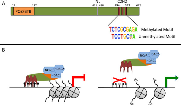
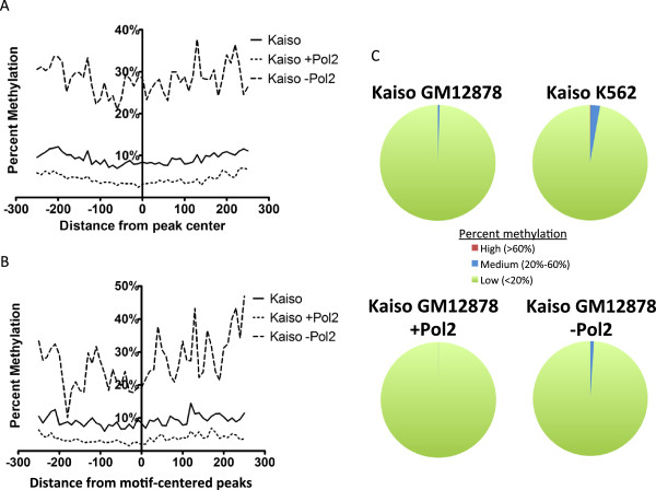
Background: DNA methylation and repressive histone modifications cooperate to silence promoters. One mechanism by which regions of methylated DNA could acquire repressive histone modifications is via methyl DNA-binding transcription factors. The zinc finger protein ZBTB33 (also known as Kaiso) has been shown in vitro to bind preferentially to methylated DNA and to interact with the SMRT/NCoR histone deacetylase complexes. We have performed bioinformatic analyses of Kaiso ChIP-seq and DNA methylation datasets to test a model whereby binding of Kaiso to methylated CpGs leads to loss of acetylated histones at target promoters.

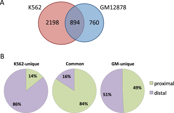
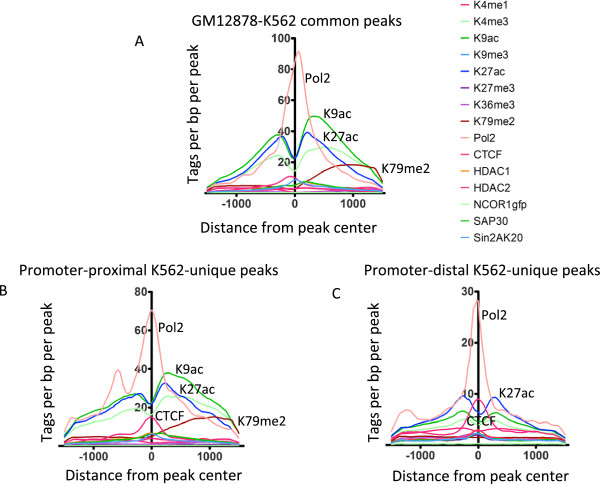
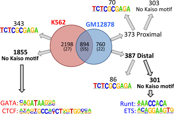
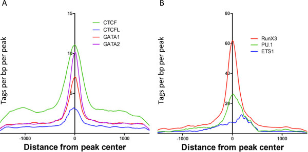
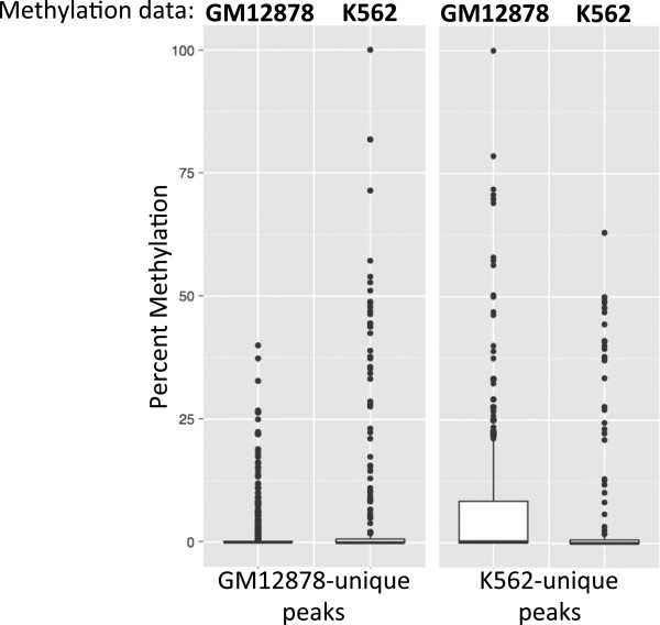
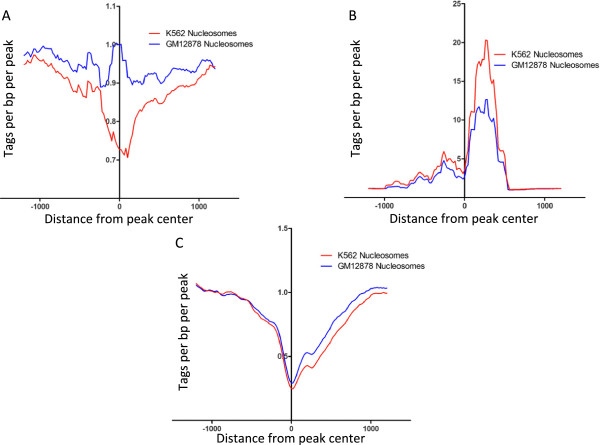
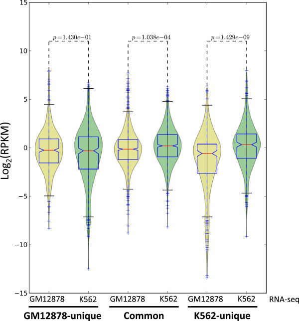
Results: Our results suggest that, contrary to expectations, Kaiso does not bind to methylated DNA in vivo but instead binds to highly active promoters that are marked with high levels of acetylated histones. In addition, our studies suggest that DNA methylation and nucleosome occupancy patterns restrict access of Kaiso to potential binding sites and influence cell type-specific binding.

Conclusions: We propose a new model for the genome-wide binding and function of Kaiso whereby Kaiso binds to unmethylated regulatory regions and contributes to the active state of target promoters.


Figures

Figure 1
Figure 1
Structure and function of Kaiso. (A) Kaiso is a 672 amino acid protein, which includes an N-terminal POZ/BTB domain required for protein-protein interactions, a nuclear localization signal, and three tandem C2H2 zinc finger domains responsible for DNA binding; numbers represent the amino acid borders of each domain. Shown below the zinc finger domains are the two motifs to which Kaiso has been shown to bind in vitro. (B) The current model for Kaiso’s activity at promoters: in the left figure, Kaiso’s 3 C2H2 zinc finger domains recognize and bind methylated DNA, recruiting the NCoR histone deacetylation complex to the region and causing the loss of active chromatin marks and the repression of the adjacent promoter. The figure on the right shows an unmethylated promoter, which is not recognized by Kaiso. As a result, the NCoR histone deacetylation complex is not recruited to the region and surrounding histones remain acetylated allowing for expression of the promoter.
Figure 2
Figure 2
Identification of high-confidence Kaiso peaks in GM12878 cells. The set of 12,543 Kaiso peaks identified in GM12878 cells using the merged replicate datasets (solid line) and the set of 1,648 high-confidence Kaiso peaks (dashed line) is plotted as tag height versus ranked peak number. The inset shows a Venn diagram of the overlap between the two Kaiso ChIP-seq replicates in GM12878 cells. The total number of peaks in each replicate is represented in parentheses; 1,648 high-confidence peaks were found to be present in both replicates.
Figure 3
Figure 3
Kaiso binds to GC-rich promoters that are also bound by Pol2. (A) Shown is the percentage of Kaiso peaks in GM12878 cells located within 1 kilobase of a Refseq transcription start site (proximal, blue), and those located further than 1 kilobase away from a transcription start site (distal, red). (B) Shown is the percentage of Kaiso peaks in GM12878 cells that are found within CpG islands. (C) Shown is the percentage of Kaiso peaks in GM12878 cells that overlap the top-ranked set of Pol2 peaks (see Additional file 2).
Figure 4
Figure 4
Epigenomic profiles of Kaiso binding sites in GM12878 cells. The density of ChIP-seq tags for several histone modifications and transcription factors is plotted relative to subsets of Kaiso binding sites in GM12878; (A) all high-confidence Kaiso peaks, (B) high-confidence Kaiso peaks that overlap with top-ranked Pol2 peaks, (C) high-confidence Kaiso peaks that do not overlap with top-ranked Pol2 peaks.
Figure 5
Figure 5
DNA methylation analysis of Kaiso peaks. (A) The average percent DNA methylation in the sequences surrounding the Kaiso binding sites (centered on the middle of the peak) was determined and plotted across all high-confidence Kaiso peaks (solid line), high-confidence Kaiso peaks overlapping Pol2 (short dashed line), and high-confidence Kaiso peaks not overlapping Pol2 (long dashed line). (B) A similar analysis was performed as in panel (A) except that only the subsets of high-confidence Kaiso peaks containing the 10 bp Kaiso motif were used and the regions were centered on the Kaiso motif. (C) Pie charts depicting the methylation percentage of all CGCG motifs in various sets of Kaiso peaks.
Figure 6
Figure 6
Characterization of cell type-specific Kaiso binding sites. (A) High-confidence Kaiso peaks in GM12878 and K562 were overlapped to identify common peaks and cell type-specific peaks between the two datasets. (B) The common and cell type-specific Kaiso peaks were analyzed for their position relative to the start site of transcription of the set of Refseq genes; sites within +/- 1 kb of a start site were classified as promoter proximal whereas all other sites were classified as promoter distal.
Figure 7
Figure 7
Epigenomic profiles of Kaiso binding sites in K562 cells. The density of ChIP-seq tags for several histone modifications and transcription factors is plotted relative to subsets of Kaiso binding sites in K562 cells; (A) peaks common between K562 and GM12878 cells, (B) promoter proximal peaks unique to K562, (C) promoter distal peaks unique to K562.
Figure 8
Figure 8
Motif analysis of cell type-specific Kaiso binding sites. Kaiso peaks from GM12878 and K562 were compared to identify common and cell type-specific binding sites (the median tag height is shown in parentheses). GM12878- and K562-unique peaks were divided into two sets of peaks, those that contain and those that lack a Kaiso motif; GM12878-unique peaks were further separated into promoter proximal and promoter distal sets. A motif analysis was then performed for each set of peaks, identifying motifs for the GATA and CTCF families of transcription factors in the K562-unique peaks and motifs for the ETS and Runt families of transcription factors in distal GM12878 peaks (see also Additional file 8).
Figure 9
Figure 9
Testing bioinformatic predictions of transcription factor co-localization. The density of ChIP-seq tags for the factors identified by motif analysis in Figure 8 were plotted relative to (A) non-motif containing K562 Kaiso peaks, and (B) promoter distal, non-motif containing GM12878 Kaiso peaks.
Figure 10
Figure 10
Methylation of cell type-specific CGCG motifs in different cell lines. The percent methylation of individual CGCG motifs within GM12878-unique and K562-unique peaks was calculated using GM12878 WGBS and RRBS data and K562 RRBS data, and graphed as box plots showing differences in methylation between cell types.
Figure 11
Figure 11
Nucleosome density of cell type-specific Kaiso peaks in GM12878 and K562. Nucleosome positioning data from MNase-seq was used and plotted as the average density of nucleosomes relative to Kaiso peaks in A) GM12878, B) K562, and C) common sites.
Figure 12
Figure 12
Expression of cell type-specific Kaiso-bound promoters in GM12878 and K562. RPKM values from RNA-seq experiments in GM12878 and K562 cells were used to create box plots showing expression of cell type-specific and common Kaiso-bound genes.

References

    1. Jones PA. Functions of DNA methylation: islands, start sites, gene bodies and beyond. Nat Rev Genet. 2012;13:484–492. doi: 10.1038/nrg3230. - DOI - PubMed
    1. Li G, Reinberg D. Chromatin higher-order structures and gene regulation. Curr Opin Genet Dev. 2011;21:175–186. doi: 10.1016/j.gde.2011.01.022. - DOI - PMC - PubMed
    1. Statham AL, Robinson MD, Song JZ, Coolen MW, Stirzaker C, Clark SJ. Bisulfite sequencing of chromatin immunoprecipitated DNA (BisChIP-seq) directly informs methylation status of histone-modified DNA. Genome Res. 2012;22:1120–1127. doi: 10.1101/gr.132076.111. - DOI - PMC - PubMed
    1. Brinkman AB, Gu H, Bartels SJJ, Zhang Y, Matarese F, Simmer F, Marks H, Bock C, Gnirke A, Meissner A, Stunnenberg HG. Sequential ChIP-bisulfite sequencing enables direct genome-scale investigation of chromatin and DNA methylation cross-talk. Genome Res. 2012;22:1128–1138. doi: 10.1101/gr.133728.111. - DOI - PMC - PubMed
    1. Komashko VM, Farnham PJ. 5-azacytidine treatment reorganizes genomic histone modification patterns. Epigenetics. 2010;5:229–240. doi: 10.4161/epi.5.3.11409. - DOI - PubMed