Review
. 2013 Sep;8(4):840-56.
doi: 10.1007/s11481-013-9470-8. Epub 2013 May 22.

Drainage of cells and soluble antigen from the CNS to regional lymph nodes

Affiliations
Review

Drainage of cells and soluble antigen from the CNS to regional lymph nodes

Jon D Laman et al. J Neuroimmune Pharmacol. 2013 Sep.

Abstract

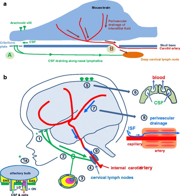
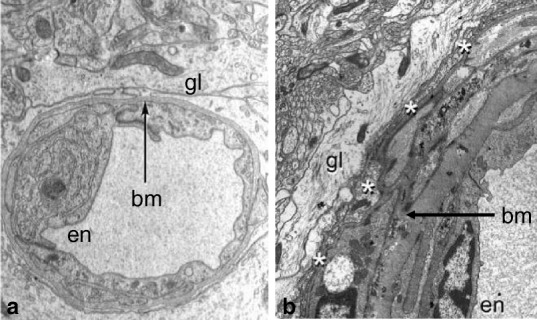
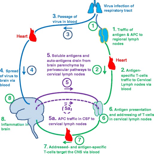
Despite the absence of conventional lymphatics, there is efficient drainage of both cerebrospinal fluid (CSF) and interstitial fluid (ISF) from the CNS to regional lymph nodes. CSF drains from the subarachnoid space by channels that pass through the cribriform plate of the ethmoid bone to the nasal mucosa and cervical lymph nodes in animals and in humans; antigen presenting cells (APC) migrate along this pathway to lymph nodes. ISF and solutes drain from the brain parenchyma to cervical lymph nodes by a separate route along 100-150 nm wide basement membranes in the walls of cerebral capillaries and arteries. This pathway is too narrow for the migration of APC so it is unlikely that APC traffic directly from brain parenchyma to lymph nodes by this route. We present a model for the pivotal involvement of regional lymph nodes in immunological reactions of the CNS. The role of regional lymph nodes in immune reactions of the CNS in virus infections, the remote influence of the gut microbiota, multiple sclerosis and stroke are discussed. Evidence is presented for the role of cervical lymph nodes in the induction of tolerance and its influence on neuroimmunological reactions. We look to the future by examining how nanoparticle technology will enhance our understanding of CNS-lymph node connections and by reviewing the implications of lymphatic drainage of the brain for diagnosis and therapy of diseases of the CNS ranging from neuroimmunological disorders to dementias. Finally, we review the challenges and opportunities for progress in CNS-lymph node interactions and their involvement in disease processes.


Figures

Fig. 1
Fig. 1
Pathways for the drainage of fluid, solutes and cells from the brain to cervical lymph nodes in rodents and humans. Two of the major fluids associated with the CNS are: (i) CSF in the ventricles and subarachnoid space, and (ii) interstitial fluid (ISF) in the brain parenchyma. CSF and ISF have separate drainage pathways. a Lymphatic drainage of the rodent brain. In rodents, a large proportion of CSF drains via the cribriform plate and nasal mucosa (A) to deep cervical lymph nodes. Arachnoid villi are very small. ISF and solutes drain from brain parenchyma along 100–150 nm-thick basement membranes in the walls of capillaries and arteries to cervical lymph nodes (B). b Lymphatic drainage of the human brain. Lymphatic drainage of CSF. CSF drains from the subarachnoid space through the cribriform plate into nasal lymphatics (1). An enlarged view of the cribriform plate (1a) shows how direct channels for the drainage of CSF from the subarachnoid space (SAS) pass alongside branches of olfactory nerves (ON) into the nasal mucosa. This pathway allows drainage of CSF and antigen presenting cells (see pink cell with blue nucleus in the drainage pathway). (2) Conventional lymphatic vessels carry fluid and cells (3) to cervical lymph nodes (4). CSF also drains into the blood through arachnoid villi and granulations (5) by macromolecular transport (6) through venous endothelial cells. Lymphatic drainage of Interstitial Fluid (ISF). ISF drains from the brain parenchyma along perivascular pathways (7); ISF and solutes diffuse through extracellular spaces of the brain and (8) enter 100–150 nm thick basement membranes in the walls of capillaries and the tunica media of arteries to drain from the brain, depicted as a blue line in (8). Fluid and solutes draining along the walls of cerebral arteries (7) pass into the wall of the internal carotid artery in the neck from which they drain to cervical lymph nodes (9). Modified from (Laman and Weller 2012)
Fig. 2
Fig. 2
Basement membranes in the walls of a human capillary and artery. (a) Capillary: the lumen is surrounded by endothelium (en) and the basement membrane (bm) encompasses the abluminal surface of the endothelium, separating it from the perivascular glia (gl). X 14000 (b) Artery wall: the endothelium (en) surrounds the lumen and basement membrane (bm) is interposed between the smooth muscle cells in the tunica media. A thin-layer of leptomeningeal cells (white asterisks) separates the perivascular glia (gl) from the artery wall. Basement membranes in the walls of capillaries and arteries are the conduits for perivascular lymphatic drainage of fluid and solutes from the CNS parenchyma as shown by tracer studies and by the distribution of Aβ in cerebral amyloid angiopathy in humans (Carare et al. 2013). X 8400 Reproduced with permission from (a) (Preston et al. 2003) (b) (Zhang et al. 1990)
Fig. 3
Fig. 3
Generation of immune and autoimmune reactions in the brain. Foreign organisms enter the body via exposed surfaces such as skin, respiratory tract and gut. 1) Antigen and antigen presenting cells from those organs traffic to regional lymph nodes and stimulate the production of antigen specific T lymphocytes that, 2) traffic to cervical lymph nodes and other groups of lymph nodes via the blood. 3 &4) organisms such as viruses reach the brain via blood. 5) Soluble viral antigens and autoantigens drain along perivascular pathways to cervical lymph nodes and 5a) antigen presenting cells (APC) in the CSF also traffic to cervical lymph nodes via CSF drainage pathways. 6) Soluble antigens and autoantigens are presented to antigen-specific T lymphocytes in the cervical lymph nodes and T cells are addressed through the induction of integrins to target the brain. 7) Integrin specific, antigen specific T lymphocytes enter the blood and traffic to the brain and induce inflammation (8)

References

    1. Abbott NJ. Evidence for bulk flow of brain interstitial fluid: significance for physiology and pathology. Neurochem Int. 2004;45:545–552. doi: 10.1016/j.neuint.2003.11.006. - DOI - PubMed
    1. Andersson PB, Perry VH, Gordon S. Intracerebral injection of proinflammatory cytokines or leukocyte chemotaxins induces minimal myelomonocytic cell recruitment to the parenchyma of the central nervous system. J Exp Med. 1992;176:255–259. doi: 10.1084/jem.176.1.255. - DOI - PMC - PubMed
    1. Bailey SL, Schreiner B, McMahon EJ, Miller SD. CNS myeloid DCs presenting endogenous myelin peptides ‘preferentially’ polarize CD4+ T(H)-17 cells in relapsing EAE. Nat Immunol. 2007;8(2):172–180. doi: 10.1038/ni1430. - DOI - PubMed
    1. Barua NU, Bienemann AS, Hesketh S, Wyatt MJ, Castrique E, Love S, Gill SS. Intrastriatal convection-enhanced delivery results in widespread perivascular distribution in a pre-clinical model. Fluids Barriers CNS. 2012;9:2. doi: 10.1186/2045-8118-9-2. - DOI - PMC - PubMed
    1. Bechmann I, Galea I, Perry VH. What is the blood–brain barrier (not)? Trends Immunol. 2007;28:5–11. doi: 10.1016/j.it.2006.11.007. - DOI - PubMed

Publication types

MeSH terms

LinkOut - more resources