Review
. 2013 Jul;6(4):666-77.
doi: 10.1038/mi.2013.30. Epub 2013 May 22.

Microfold (M) cells: important immunosurveillance posts in the intestinal epithelium

Affiliations
Review

Microfold (M) cells: important immunosurveillance posts in the intestinal epithelium

N A Mabbott et al. Mucosal Immunol. 2013 Jul.

Abstract

The transcytosis of antigens across the gut epithelium by microfold cells (M cells) is important for the induction of efficient immune responses to some mucosal antigens in Peyer's patches. Recently, substantial progress has been made in our understanding of the factors that influence the development and function of M cells. This review highlights these important advances, with particular emphasis on: the host genes which control the functional maturation of M cells; how this knowledge has led to the rapid advance in our understanding of M-cell biology in the steady state and during aging; molecules expressed on M cells which appear to be used as "immunosurveillance" receptors to sample pathogenic microorganisms in the gut; how certain pathogens appear to exploit M cells to infect the host; and finally how this knowledge has been used to specifically target antigens to M cells to attempt to improve the efficacy of mucosal vaccines.


Figures

Figure 1
Figure 1
The morphological features of M cells. (a) Cartoon illustrating the morphological features of M cells. Note the lack of microvilli and basolateral pocket containing a mononuclear phagocyte and a lymphocyte. (b) Whole-mount immunohistochemical analysis of GP2+ mature M cells (green) in the FAE of a mouse Peyer’s patch. Note the regular distribution of the M cells across the FAE. Note also that in the steady-state M cells are restricted to the FAE and are mostly absent from the surrounding villi. Tissue counterstained to detect f-actin (blue). V, villi. (c) Scanning electron micrograph of the apical surface of a M-cell. Note characteristic lack of/blunted microvilli.
Figure 2
Figure 2
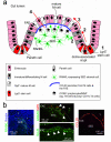
M cells differentiate from Lgr5+ stem cells in the crypts in a RANKL- and Spi-B dependent manner. (a, 1) All epithelial cell lineages, including M cells, develop from Lgr5+ intestinal epithelial stem cells within the crypts. (a, 2) In the intestine RANKL is selectively expressed by the subepithelial stromal cells beneath the FAE, . RANKL controls the differentiation of Lgr5+ stem cell-derived RANK-expressing enterocytes, into M cells, . (a, 3) RANKL also induces the expression of the ETS family transcription factor Spi-B. The subsequent differentiation of the ANXA5+, MARCKS-like protein 1+ immature M cells into functionally mature GP2+, TNFAIP2+ and CCL9+ M cells is regulated by intrinsic expression of Spi-B, , . (a, 4) The chemokine CCL20 is specifically expressed by the FAE, and in Peyer’s patches it mediates the chemoattraction of CCR6-expressing lymphocytes and leukocytes towards the FAE. When CCL20-CCR6 signalling is impeded, M-cell maturation is likewise impeded-. Data suggest that a unique subset of CCR6hiCD11cint B cells specifically migrates towards FAE-derived CCL20 and promotes M-cell differentiation. However, since M-cell maturation can be induced in an in vitro “gut organoid” model system containing only epithelial cell elements, it plausible that these CCL20-CCR6-mediated lympho-epithelial interactions most likely provide accessory factors that enhance M-cell differentiation. (b) Immunohistochemical analysis of the distribution of RANKL (green, left-hand panel), ANXA5, GP2 (green and red, respectively, upper-middle panel), Spi-B (green, lower-middle panel) and CCL20 (red, right-hand panel) in mouse Peyer’s patches. Dotted lines indicate the boundary of the FAE. Arrows indicate positively immunostained M cells. V, villi.
Figure 3
Figure 3
Retrospective comparison of chemokine gene expression by M cells and the FAE. (a) Heat map showing the mean expression of profile of multiple chemokine-encoding probe sets in samples of Peyer’s patch (PP) M cells, cholera-toxin-induced (CT-stim.) villous M cells and ileum intestinal epithelial cells (GSE7838), FAE and M cells and ileum (GSE13908). These data were performed on Affymetrix MOE430_2 mouse genome expression arrays. Each column represents the mean probe set intensity (log2) for all samples from each source (n = 2-4). Significant differences between groups were sought by ANOVA. P values for those genes which were expressed significantly (P < 0.05) within the FAE and by M cells at levels > 2.0 fold when compared to the villous epithelium are indicated. (b) Effect of RANKL-stimulation on the expression of chemokine-encoding genes in the villous epithelium. These data (GSE37861) were performed on Affymetrix mouse gene 1.0 ST expression arrays (equivalent chemokine-related probe sets are shown). Each column represents the mean probe set intensity (log2) for all samples from each source (n = 3). P values for those genes which were significantly upregulated > 2.0 fold at d 3 after RANKL-treatments when compared to controls (d 0) are indicated. Representative probe set are shown when multiple probe sets for a gene were present on the arrays. NS, not significant.
Figure 4
Figure 4
Potential routes of lumenal antigen sampling in the intestine by epithelial cell subsets and MNP. (1) Sampling of gut lumenal microorganisms and macromolecules via M-cell-mediated transcytosis. (2) Although the incidence is rare in the steady-state, certain inflammatory stimuli appear to recruit MNP from the lamina propria where they insert their dendrites through the tight junctions between enterocytes to directly sample the luminal contents, . (3) Another study has reported that MNP may even sample lumenal antigens by extending their dendrites directly through “transcellular pores” in the M cells themselves. (4) Mucin secreting goblet cells can also function as passages for the delivery of low molecular weight soluble antigens to CD103+ MNP in the lamina propria. (5) Ultrastructural analysis suggests that FAE enterocytes can also acquire certain gut lumenal antigens, and exocytose the intravascular contents of their large late endosomes into the extracellular space of the SED where they are acquired by MNP.

References

    1. Lorenz RG, Newberry RD. Isolated lymphoid follicles can function as sites for induction of mucosal immune responses. Ann. New York Acad. Sci. 2004;1029:44–57. - PubMed
    1. Kanaya T, et al. The Ets transcription factor Spi-B is essential for the differentiation of intestinal microfold cells. Nat. Immunol. 2012;13:729–736. - PMC - PubMed
    1. Tahoun A, et al. Salmonella transforms follicle-associated epithelial cells into M cells to promote intestinal invasion. Cell Host & Microbe. 2012;12:645–666. - PubMed
    1. Mabbott NA, Baillie JC, Hume DA, Freeman TC. Meta-analysis of co-expressed gene signatures in mouse leukocyte populations. Immunobiology. 2010;215:724–736. - PubMed
    1. Bradford BM, Sester D, Hume DA, Mabbott NA. Defining the anatomical localisation of subsets of the murine mononuclear phagocyte system using integrin alpha X (ITGAX) and colony stimulating factor 1 receptor (CSF1-R) expression fails to discriminate dendritic cells from macrophages. Immunobiology. 2011;216:1228–1237. - PubMed

Publication types

MeSH terms