. 2013:4:1886.
doi: 10.1038/ncomms2877.

Distinct bone marrow-derived and tissue-resident macrophage lineages proliferate at key stages during inflammation

Affiliations

Distinct bone marrow-derived and tissue-resident macrophage lineages proliferate at key stages during inflammation

Luke C Davies et al. Nat Commun. 2013.

Abstract

The general paradigm is that monocytes are recruited to sites of inflammation and terminally differentiate into macrophages. There has been no demonstration of proliferation of peripherally-derived inflammatory macrophages under physiological conditions. Here we show that proliferation of both bone marrow-derived inflammatory and tissue-resident macrophage lineage branches is a key feature of the inflammatory process with major implications for the mechanisms underlying recovery from inflammation. Both macrophage lineage branches are dependent on M-CSF during inflammation, and thus the potential for therapeutic interventions is marked. Furthermore, these observations are independent of Th2 immunity. These studies indicate that the proliferation of distinct macrophage populations provides a general mechanism for macrophage expansion at key stages during inflammation, and separate control mechanisms are implicated.


Figures
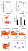
Figure 1
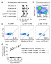
Figure 1. Kinetics of acute zymosan induced peritonitis and appearance of Ly-6B expressing inflammatory macrophages
a) Graphs showing the numbers of neutrophils and eosinophils (left panel) and MØ (both total monocyte/MØ and Res MØ) MØ (right panel) present at the indicated times after i.p. administration of 2×10⁶ zymosan particles. Data represents mean±SEM of 3-5 individual mice per time point, taken from one of two similar experiments. b) Representative flow-cytometric profiles showing Ly-6B+ Inf MØ, identified as previously described, by gating on CD11bhighF4/80low-high cells (excluding eosinophils), are clearly distinct from Tim4high Res MØ. Gates denote Ly-6B+ and Ly-6B Inf MØ populations. c) Approximately a fifth of cells classified as Inf MØ are Ly-6B+ and are present for several weeks after induction of a mild inflammatory reaction. Data is taken from the animals represented in (A) above and presented as mean±SEM. d) Representative flow-cytometric profiles (gated on F4/80low-highCD11b+ monocytes and MØ) taken from mice 72 hours after induction of peritonitis with zymosan (both 2×10⁶ and 2×10⁷ particles) and thioglycollate broth, as indicated. The higher dose of zymosan resulted in similar MØ populations, but with substantially reduced recoverable numbers of Res MØ when compared to the low dose, as expected,. In contrast, thioglycollate broth resulted in a near complete depletion of Res MØ and no evident Ly-6B+ Inf MØ population at this time. The presence of both Ly-6B+ monocytes and Ly-6B+ Inf MØ populations are indicated. Flow-cytometric plots are representative of at least 3 mice per group from one of two similar experiments. e) The Ly-6B expressing MØ were gated as indicated (left panel) and purified (>90%) by flow-cytometric cell-sorting and analysed on cytospins stained with eosin and methylene blue. Cells were pooled from 6 week old female C57BL/6 mice 7 days after i.p. zymosan (2×10⁶ particles).
Figure 2
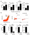
Figure 2. Both Ly-6B+ and Ly-6Binflammatory macrophages are derived from the bone marrow
Partial BM chimeras were generated as previously described , after shielding the peritoneal cavity, and then were left to recover for 6 weeks. a) Analysis of blood myeloid cell chimerism one day before induction of peritonitis shows a stable chimerism in all experimental animals. Each line links the data from an individual mouse. b) Gating strategy for the identification of Res and Inf MØ (both Ly-6B+ and Ly-6B) 72 hours after the induction of peritonitis by i.p. administration of 2×10⁶ zymosan particles. The plot has been pre-gated on CD11bhighF4/80low-high cells as previously reported. c) Representative flow-cytometric analysis of the indicated MØ subsets to determine the level of chimerism between CD45.1 recipient cells and CD45.2 donor cells. d) Graphical representation of the chimerism in all of the experimental animals correlating the chimerism of the 3 peritoneal MØ subsets with that of Ly-6Chigh blood monocytes within the same animal. Data were analyzed by linear regression, and r2 values are indicated alongside significance testing for a non-zero slope. Each symbol represents an individual irradiation chimera.
Figure 3
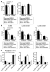
Figure 3. Ly-6B expressing inflammatory macrophages actively dividein situ
a) The plots show total Inf MØ populations (left), Ly-6B+ cells (middle) and their Ly-6B counterparts (right) 72 hours after the induction of peritonitis with 2×10⁶ zymosan particles. As previously reported there is only limited evidence of proliferation in the total Inf MØ population (left and ). However, when subdivided based on Ly-6B expression there was a clear enrichment of cells in the S,G2 and M stages of cell cycle amongst the Ly-6B expressing cells (middle) and notable lack of proliferating cells within the Ly-6B population (right). Data are derived from individual mice and are representative of at least 3 experiments with 6-7 week old C57BL/6 female mice and typically 3-5 mice per experiment. b) The levels of proliferation within the two populations of Inf MØ in the same experiment shown above (A) was measured by the proportion of cells with >2N DNA content (left), >2N DNA content with Ki67 expression (‘SG2/M’, middle), phospho-Histone H3+ phase of mitosis (‘pHH3+’; right). Data is expressed as mean±SEM of 5 mice per group (7 week old female C57BL/6) and is representative of at least 3 experiments. Data were analyzed by a paired t-test.
Figure 4
Figure 4. M-CSF is required for tissue resident macrophage proliferation
a) Data showing proliferation of Res MØ (gated by their F4/80highCD11bhigh phenotype), 24 hours after i.p. administration of 0.4 μg of recombinant M-CSF (0.4) or carrier control (0). Data shown represents mean±SEM 10 mice per group (7 week old female C57BL/6) pooled from 2 identical experiments. Data were analyzed by a Student’s t-test. b) Representative flow cytometric plots showing the identification of Res MØ in a similar way as previously described 48 hours after the i.p. injection of 2×10⁶ zymosan particles in conjunction with 0.5 mg of either anti-M-CSF (clone 5A1) or and rat IgGisotype control (as indicated). Percentages shown are mean values from the analyses of groups of mice (shown below). c) Measurement of proliferation in Res MØ 48 hours after i.p. injection of 2×10⁶ zymosan particles in conjunction with 0.5 mg of either anti-M-CSF (clone 5A1) or and rat IgGisotype control. Data shown represents mean±SEM 5 mice per group (7 week old female C57BL/6) from one of 2 similar experiments. Data were analyzed by a Student’s t-test.
Figure 5
Figure 5. Ly-6B expressing inflammatory macrophagesare M-CSF-dependent
Inf MØ were quantified 48 hours after acute peritonitis induced with 2×10⁶ zymosan particles in conjunction with 0.5 mg of either anti-M-CSF (clone 5A1) or and rat IgGisotype control. Neutralization of M-CSF results in a substantial drop in total Inf MØ numbers (left), which is caused by a near complete loss of Ly-6B+ Inf MØ (middle) and substantial drop in Ly-6B Inf MØ (right). Data shown represents mean±SEM of two independent experiments conducted with 7-8 week old female C57BL/6 mice (n=5 per group) and one with 9-12 week old female 129S6/SvEv (n=3-6 per group). Data was analyzed by Two-way ANOVA with the significance of the effects of both M-CSF and strain of mouse indicated.
Figure 6
Figure 6. Lack of an essential role for IL-4/IL-4Rα for macrophage proliferation during acute inflammation
a) Measurement of proliferation in Res MØ 48 hours after i.p. injection of 2×10⁶ zymosan particles in conjunction with 0.5 mg of either anti-IL-4 (clone 11B11) or and rat IgGisotype control. The left graph shows the proportion of Res MØ in the S, G2 and M phases of cell cycle whereas the right graph shows those cells in pHH3+ definitive stages of mitosis. b) Analysis of proliferation of Inf MØ subsets in mice injected i.p. 48 hours earlier with 2×10⁶ zymosan particles in conjunction with 0.5 mg of either anti-IL-4 (clone 11B11) or and rat IgGisotype control. Data shown for (A) and (B) represent mean±SEM from two-independent experiments (7 week old female C57BL/6 n=5 mice per group, and 6-9 week old female 129S6 mice n=3-6 mice per group). Data were analyzed by a Two-way ANOVA with the significance of the effects of IL-4 neutralization and mouse strain indicated. c) The number and proliferation of the Res and Inf MØ subsets were examined 72 hours after induction of peritonitis with 2×10⁶ zymosan particles in wild type and IL-4Rα deficient mice on the C57BL/6 background. Data shown represents mean±SEM of 6 week old female mice (n=10 per group).

References

    1. Geissmann F, Manz MG, Jung S, Sieweke MH, Merad M, Ley K. Development of monocytes, macrophages, and dendritic cells. Science. 2010;327:656–661. - PMC - PubMed
    1. Gordon S, Taylor PR. Monocyte and macrophage heterogeneity. Nat Rev Immunol. 2005;5:953–964. - PubMed
    1. Passlick B, Flieger D, Ziegler-Heitbrock HW. Identification and characterization of a novel monocyte subpopulation in human peripheral blood. Blood. 1989;74:2527–2534. - PubMed
    1. Geissmann F, Jung S, Littman DR. Blood monocytes consist of two principal subsets with distinct migratory properties. Immunity. 2003;19:71–82. - PubMed
    1. Sunderkotter C, Nikolic T, Dillon MJ, et al. Subpopulations of mouse blood monocytes differ in maturation stage and inflammatory response. J Immunol. 2004;172:4410–4417. - PubMed

Publication types