. 2013 Oct;6(5):762-71.
doi: 10.1007/s12265-013-9473-0. Epub 2013 May 22.

Prospective validation that vulnerable plaque associated with major adverse outcomes have larger plaque volume, less dense calcium, and more non-calcified plaque by quantitative, three-dimensional measurements using intravascular ultrasound with radiofrequency backscatter analysis : results from the ATLANTA I Study

Affiliations

Prospective validation that vulnerable plaque associated with major adverse outcomes have larger plaque volume, less dense calcium, and more non-calcified plaque by quantitative, three-dimensional measurements using intravascular ultrasound with radiofrequency backscatter analysis : results from the ATLANTA I Study

Jesus G Vazquez-Figueroa et al. J Cardiovasc Transl Res. 2013 Oct.

Abstract

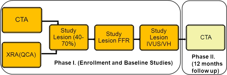
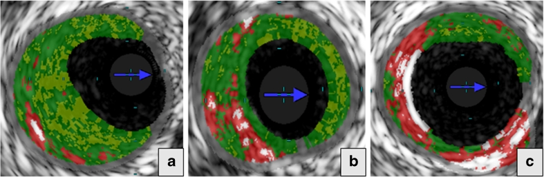
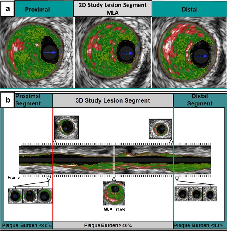
Whether quantitative, two-dimensional, and three-dimensional plaque measurements by intravascular ultrasound with radiofrequency backscatter (IVUS/VH) are different between intermediate lesions with or without major adverse cardiovascular events (MACE) is unknown. IVUS/VH-derived parameters were compared in 60 patients with an intermediate coronary lesion (40-70 %) between lesions that did or did not result in MACE over 12 months. IVUS/VH measurements were done at the site of the minimal lumen area (MLA) and on a per-plaque basis, defined by 40 % plaque burden. Pre-specified, adjudicated MACE events occurred in 5 of 60 patients (8.3 %). MACE lesions had larger plaque burden (65 % vs. 53 %, p = 0.004), less dense calcium (6.6 % vs. 14.7 %, p = 0.05), and more non-calcified plaque, mostly fibrofatty kind (17.6 % vs. 10 %, p = 0.02). Intermediate coronary lesions associated with MACE at 12 months have more plaque, less dense calcium, and more non-calcified plaque, particularly fibrofatty tissue by IVUS/VH.

Trial registration: ClinicalTrials.gov NCT00817102.


Figures

Fig. 1
Fig. 1
ATLANTA study design. Patients with intermediate lesions (40–70 %) were recruited based on XRA or CTA; each pre-specified study lesion underwent IVUS/VH and FFR. CTA was repeated at 12 months. CTA computed tomography angiography, XRA invasive X-ray coronary angiography, QCA quantitative coronary angiography, FFR fractional flow reserve, IVUS intravascular ultrasound, VH virtual histology
Fig. 2
Fig. 2
The figure represents the schematic for calculation of plaque burden for 2D study segment. 2D bi-dimensional, EEL external elastic lamina, IEL internal elastic lamina, V vessel area, L lumen area
Fig. 3
Fig. 3
Plaque composition by intravascular ultrasound with radiofrequency backscatter analysis (IVUS/VH). IVUS/VH segment is shown in the entire longitudinal section (a) and in cross-section at the minimal luminal area (MLA) frame (b)
Fig. 4
Fig. 4
Morphological lesion subtypes identified by intravascular ultrasound with radiofrequency backscatter analysis (IVUS/VH). Three plaque subtypes are shown: (a) pathological intimal thickening (PIT), (b) thick-cap fibroatheroma (VH-ThCFA), and (c) thin-cap fibroatheroma (VH-TCFA)
Fig. 5
Fig. 5
Schematic representation of 2D and 3D IVUS/VH analysis. For the 2D-IVUS/VH analysis, plaque geometrical parameters (MLD, MLA, and plaque burden) were measured in the MLA frame as well as in a frame proximal and distal to the MLA, and the values from these three consecutive frames were averaged (a). For the 3D-IVUS/VH analysis, in each subject, the entire vessel pullback from the IVUS/VH dataset was analyzed frame-by-frame, and the MLA frame was identified. The study segment was extended proximally and distally from the MLA frame until three consecutive frames had less than 40 % plaque burden (b). 2D bi-dimensional, 3D three-dimensional, IVUS intravascular ultrasound, VH virtual histology, MLA minimal lumen area
Fig. 6
Fig. 6
Significant predictors of MACE via the two-dimensional (2D; top row) and three-dimensional (3D; bottom row) IVUS/VH analyses. MACE major adverse cardiac events, 2D bi-dimensional, 3D three-dimensional, IVUS intravascular ultrasound, VH virtual histology

References

    1. Writing Group. Lloyd-Jones D, Adams RJ, et al. Heart disease and stroke statistics—2010 update: a report from the American Heart Association. Circulation. 2010;121:e46–e215. doi: 10.1161/CIRCULATIONAHA.109.192667. - DOI - PubMed
    1. Cannon CP, Braunwald E, McCabe CH, et al. Intensive versus moderate lipid lowering with statins after acute coronary syndromes. The New England Journal of Medicine. 2004;350:1495–1504. doi: 10.1056/NEJMoa040583. - DOI - PubMed
    1. Voros S. "Does imaging paint a sugar-coated picture of diabetic vessels?": plaque composition in diabetics by IVUS and CT angiography. Journal of Nuclear Cardiology. 2009;16:339–344. doi: 10.1007/s12350-009-9069-x. - DOI - PubMed
    1. Muller JE, Tofler GH, Stone PH. Circadian variation and triggers of onset of acute cardiovascular disease. Circulation. 1989;79:733–743. doi: 10.1161/01.CIR.79.4.733. - DOI - PubMed
    1. Virmani R, Burke AP, Farb A, Kolodgie FD. Pathology of the vulnerable plaque. Journal of the American College of Cardiology. 2006;47:C13–C18. doi: 10.1016/j.jacc.2005.10.065. - DOI - PubMed

Publication types

MeSH terms

Associated data