. 2014 Feb;36(1):21-30.
doi: 10.1007/s11357-013-9540-0. Epub 2013 May 22.

Skeletal muscle ATP kinetics are impaired in frail mice

Affiliations

Skeletal muscle ATP kinetics are impaired in frail mice

Ashwin Akki et al. Age (Dordr). 2014 Feb.

Abstract

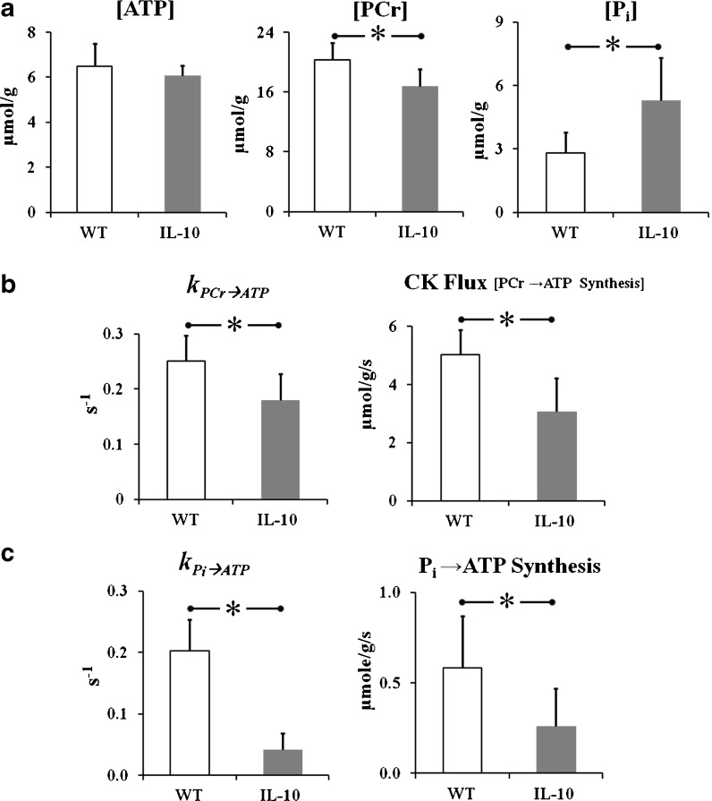
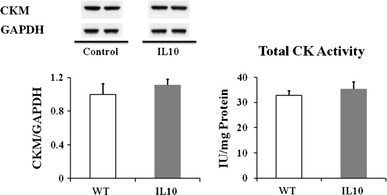
The interleukin-10 knockout mouse (IL10(tm/tm)) has been proposed as a model for human frailty, a geriatric syndrome characterized by skeletal muscle (SM) weakness, because it develops an age-related decline in SM strength compared to control (C57BL/6J) mice. Compromised energy metabolism and energy deprivation appear to play a central role in muscle weakness in metabolic myopathies and muscular dystrophies. Nonetheless, it is not known whether SM energy metabolism is altered in frailty. A combination of in vivo (31)P nuclear magnetic resonance experiments and biochemical assays was used to measure high-energy phosphate concentrations, the rate of ATP synthesis via creatine kinase (CK), the primary energy reserve reaction in SM, as well as the unidirectional rates of ATP synthesis from inorganic phosphate (Pi) in hind limb SM of 92-week-old control (n = 7) and IL10(tm/tm) (n = 6) mice. SM Phosphocreatine (20.2 ± 2.3 vs. 16.8 ± 2.3 μmol/g, control vs. IL10(tm/tm), p < 0.05), ATP flux via CK (5.0 ± 0.9 vs. 3.1 ± 1.1 μmol/g/s, p < 0.01), ATP synthesis from inorganic phosphate (Pi → ATP) (0.58 ± 0.3 vs. 0.26 ± 0.2 μmol/g/s, p < 0.05) and the free energy released from ATP hydrolysis (∆G ∼ATP) were significantly lower and [Pi] (2.8 ± 1.0 vs. 5.3 ± 2.0 μmol/g, control vs. IL10(tm/tm), p < 0.05) markedly higher in IL10(tm/tm) than in control mice. These observations demonstrate that, despite normal in vitro metabolic enzyme activities, in vivo SM ATP kinetics, high-energy phosphate levels and energy release from ATP hydrolysis are reduced and inorganic phosphate is elevated in a murine model of frailty. These observations do not prove, but are consistent with the premise, that energetic abnormalities may contribute metabolically to SM weakness in this geriatric syndrome.


Figures

Fig. 1
Fig. 1
Fully relaxed 31P MR spectra. Representative fully relaxed 31P MR spectra from the hind limb skeletal muscle of control (a) and IL10tm/tm (b) mice. Note the substantially higher inorganic phosphate (Pi) signal in the skeletal muscle of IL10tm/tm mouse
Fig. 2
Fig. 2
31P Magnetization Transfer Spectra. Representative 31P MT spectra from the hind limb skeletal muscle of control (a) and IL10tm/tm (b) mice. The spectra were acquired with saturating irradiation (thick arrows) in the control position (left spectrum in each panel) and γ-ATP position (right spectrum in each panel). The decrease in the height of PCr peak between control and γ-ATP saturation (slope of the dotted lines) is directly related to the rate of ATP synthesis through the CK reaction. A smaller decline in PCr signal with γ-ATP saturation observed in IL10tm/tm vs. control mice indicates lower ATP flux through CK in the skeletal muscle of IL10tm/tm mice
Fig. 3
Fig. 3
Skeletal Muscle Energetics. Skeletal muscle concentrations of ATP, Phosphocreatine (PCr) and Pi (a), the CK pseudo-first order rate constant (k PCr → ATP) and rate of ATP synthesis through CK (i.e., CK Flux) (b), and the pseudo-first order rate constant for ATP synthesis from Pi (k Pi → ATP) and unidirectional (Pi → ATP) rates of ATP synthesis (c) in the hind limb of control (white bars) and IL10tm/tm (gray bars) mice. The high-energy phosphate, PCr, was lower, Pi was higher and rates of ATP synthesis through both CK and from Pi, were significantly reduced in IL10tm/tm mice as compared to controls. Results are mean ± SD for n = 6–7 in each group. *p < 0.05 vs. control
Fig. 4
Fig. 4
Skeletal Muscle CK expression and activity. Skeletal muscle CKM protein expression and in vitro total CK activity in control and IL10tm/tm mice. Results expressed as mean ± SD for n = 6–7 for each group
Fig. 5
Fig. 5
Skeletal muscle catalase activity and protein carbonylation. Skeletal muscle catalase activity (left) and protein carbonylation (right) in control (“WT” white bars) and IL10tm/tm mice (“IL10”, gray bars). Catalase activity was significantly higher (*p < 0.02) and protein carbonylation trended higher (p = 0.06) in IL10 as compared to control hearts. Results expressed as mean ± SD for n = 6–7 for each group

References

    1. Allen DG, Lamb GD, Westerblad H. Skeletal muscle fatigue: cellular mechanisms. Physiol Rev. 2008;88:287–332. doi: 10.1152/physrev.00015.2007. - DOI - PubMed
    1. Balaban RS, Koretsky AP. Interpretation of 31P NMR saturation transfer experiments: what you can’t see might confuse you. Focus on "Standard magnetic resonance-based measurements of the Pi–>ATP rate do not index the rate of oxidative phosphorylation in cardiac and skeletal muscles". Am J of Physiol Cell Physiol. 2011;301:C12–15. doi: 10.1152/ajpcell.00100.2011. - DOI - PMC - PubMed
    1. Barnes PR, Kemp GJ, Taylor DJ, Radda GK. Skeletal muscle metabolism in myotonic dystrophy A 31P magnetic resonance spectroscopy study. Brain : A J of Neurol. 1997;120(Pt 10):1699–1711. doi: 10.1093/brain/120.10.1699. - DOI - PubMed
    1. Bittl JA, DeLayre J, Ingwall JS. Rate equation for creatine kinase predicts the in vivo reaction velocity: 31P NMR surface coil studies in brain, heart, and skeletal muscle of the living rat. Biochemistry. 1987;26:6083–6090. doi: 10.1021/bi00393a021. - DOI - PubMed
    1. Cline GW, Vidal-Puig AJ, Dufour S, Cadman KS, Lowell BB, Shulman GI. In vivo effects of uncoupling protein-3 gene disruption on mitochondrial energy metabolism. J Biol Chem. 2001;276:20240–20244. doi: 10.1074/jbc.M102540200. - DOI - PubMed