

Abnormal glycosphingolipid mannosylation triggers salicylic acid-mediated responses in Arabidopsis
- PMID: 23695979
- PMCID: PMC3694712
- DOI: 10.1105/tpc.113.111500
Abnormal glycosphingolipid mannosylation triggers salicylic acid-mediated responses in Arabidopsis
Abstract
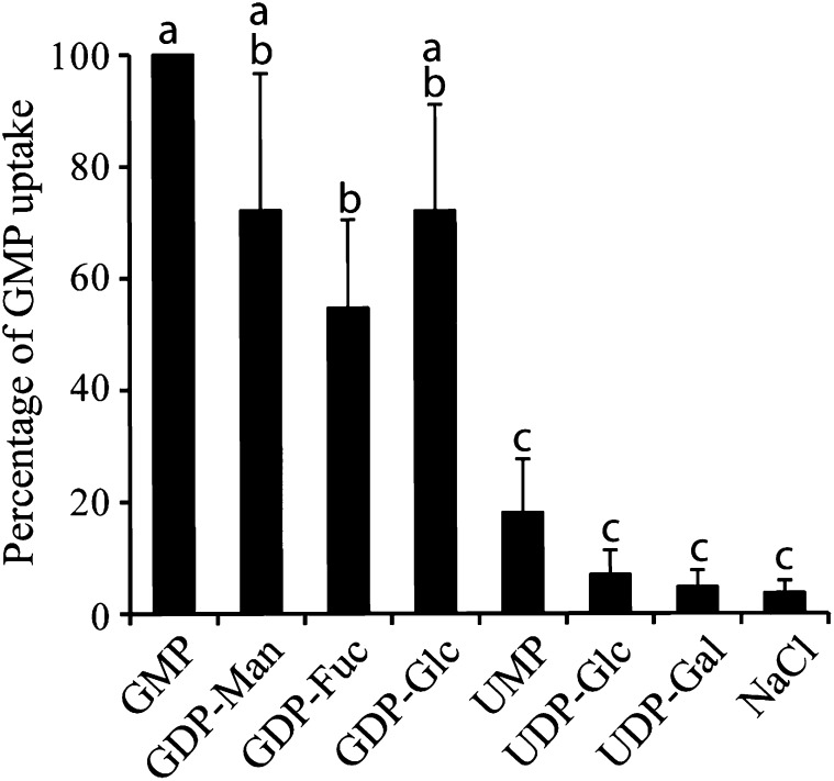
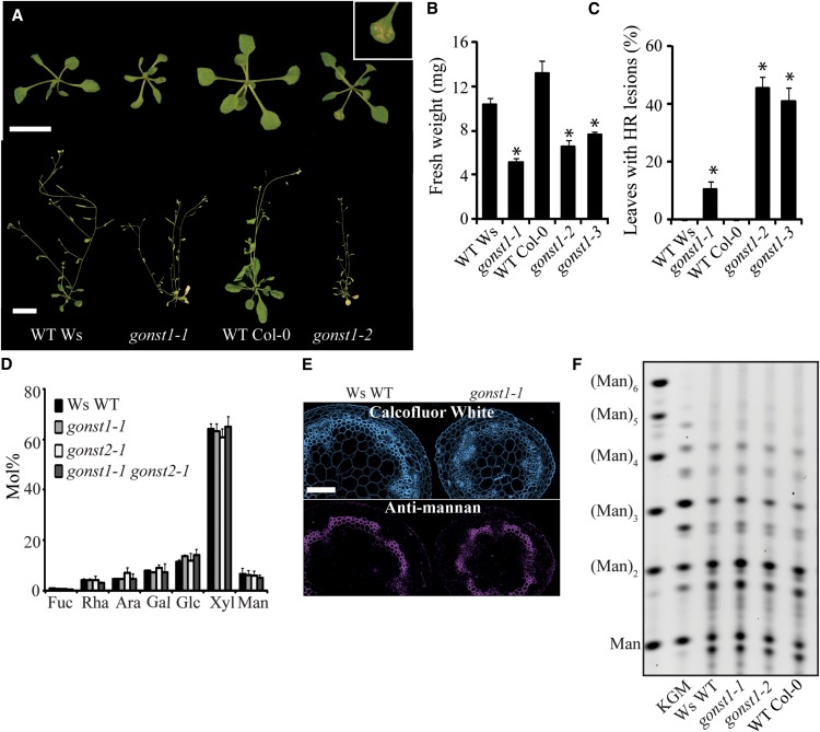
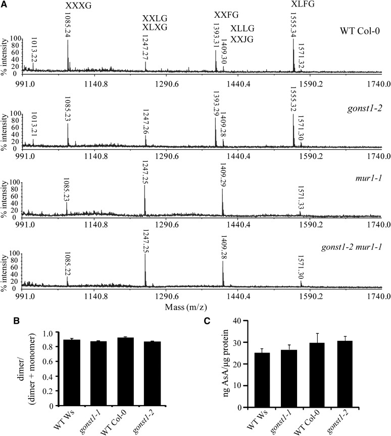
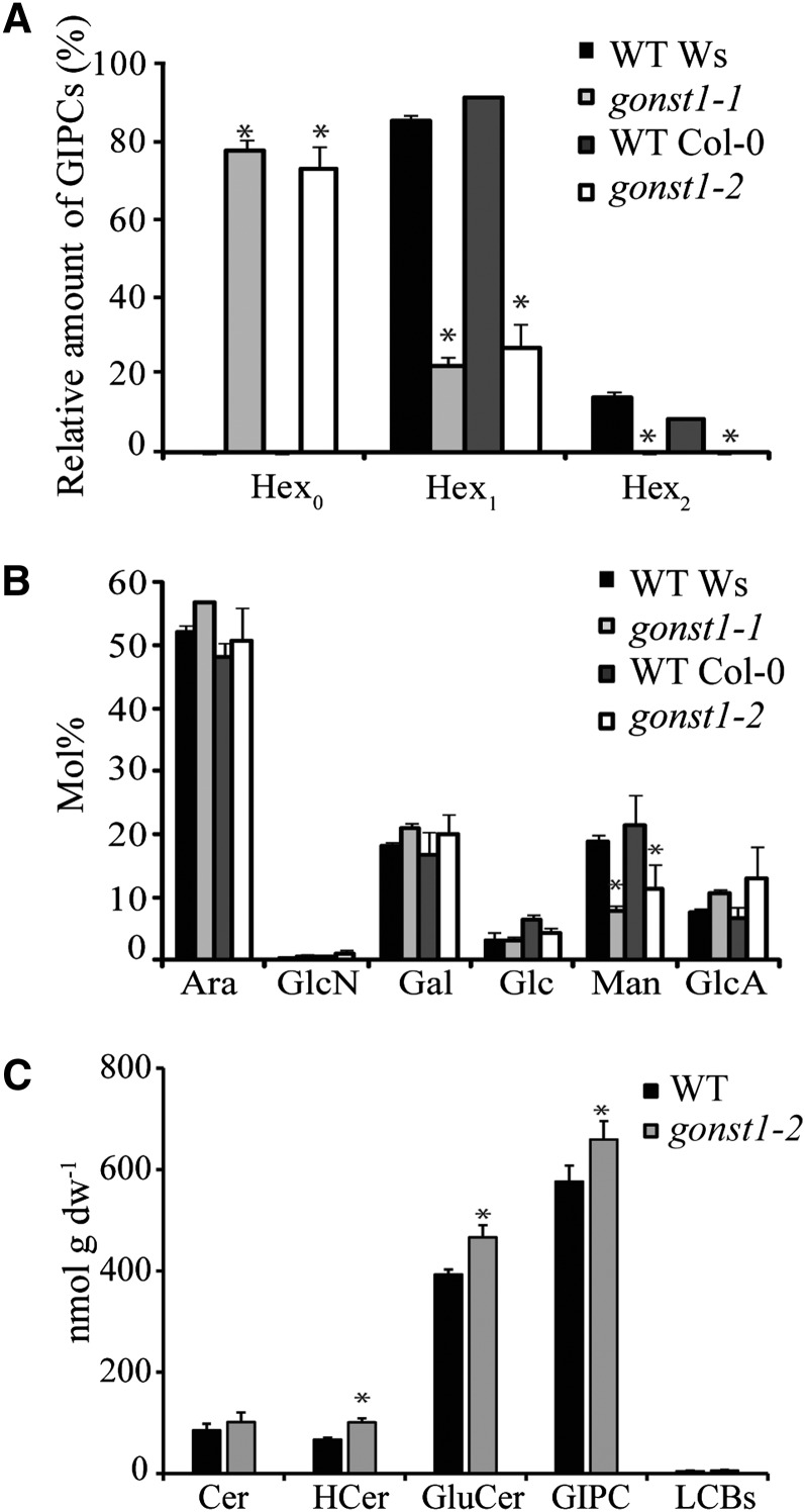
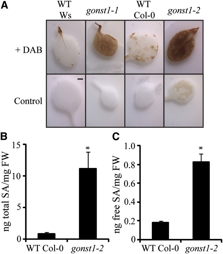
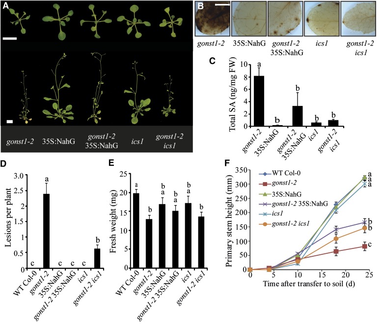
The Arabidopsis thaliana protein GOLGI-LOCALIZED NUCLEOTIDE SUGAR TRANSPORTER (GONST1) has been previously identified as a GDP-d-mannose transporter. It has been hypothesized that GONST1 provides precursors for the synthesis of cell wall polysaccharides, such as glucomannan. Here, we show that in vitro GONST1 can transport all four plant GDP-sugars. However, gonst1 mutants have no reduction in glucomannan quantity and show no detectable alterations in other cell wall polysaccharides. By contrast, we show that a class of glycosylated sphingolipids (glycosylinositol phosphoceramides [GIPCs]) contains Man and that this mannosylation is affected in gonst1. GONST1 therefore is a Golgi GDP-sugar transporter that specifically supplies GDP-Man to the Golgi lumen for GIPC synthesis. gonst1 plants have a dwarfed phenotype and a constitutive hypersensitive response with elevated salicylic acid levels. This suggests an unexpected role for GIPC sugar decorations in sphingolipid function and plant defense signaling. Additionally, we discuss these data in the context of substrate channeling within the Golgi.
Figures
Similar articles
-
Arabidopsis thaliana expresses multiple Golgi-localised nucleotide-sugar transporters related to GONST1.Mol Genet Genomics. 2004 Nov;272(4):397-410. doi: 10.1007/s00438-004-1071-z. Epub 2004 Oct 8. Mol Genet Genomics. 2004. PMID: 15480787
-
Loss of Inositol Phosphorylceramide Sphingolipid Mannosylation Induces Plant Immune Responses and Reduces Cellulose Content in Arabidopsis.Plant Cell. 2016 Dec;28(12):2991-3004. doi: 10.1105/tpc.16.00186. Epub 2016 Nov 28. Plant Cell. 2016. PMID: 27895225 Free PMC article.
-
Identification and characterization of GONST1, a golgi-localized GDP-mannose transporter in Arabidopsis.Plant Cell. 2001 Oct;13(10):2283-95. doi: 10.1105/tpc.010247. Plant Cell. 2001. PMID: 11595802 Free PMC article.
-
The inside and outside: topological issues in plant cell wall biosynthesis and the roles of nucleotide sugar transporters.Glycobiology. 2016 Sep;26(9):913-925. doi: 10.1093/glycob/cww054. Epub 2016 Aug 9. Glycobiology. 2016. PMID: 27507902 Review.
-
Gateway to the Golgi: molecular mechanisms of nucleotide sugar transporters.Curr Opin Struct Biol. 2019 Aug;57:127-134. doi: 10.1016/j.sbi.2019.03.019. Epub 2019 Apr 15. Curr Opin Struct Biol. 2019. PMID: 30999236 Free PMC article. Review.
Cited by
-
Sphingolipids: towards an integrated view of metabolism during the plant stress response.New Phytol. 2020 Jan;225(2):659-670. doi: 10.1111/nph.15997. Epub 2019 Jul 15. New Phytol. 2020. PMID: 31211869 Free PMC article. Review.
-
Knocking Down the Expression of GMPase Gene OsVTC1-1 Decreases Salt Tolerance of Rice at Seedling and Reproductive Stages.PLoS One. 2016 Dec 19;11(12):e0168650. doi: 10.1371/journal.pone.0168650. eCollection 2016. PLoS One. 2016. PMID: 27992560 Free PMC article.
-
Xylan in the Middle: Understanding Xylan Biosynthesis and Its Metabolic Dependencies Toward Improving Wood Fiber for Industrial Processing.Front Plant Sci. 2019 Feb 25;10:176. doi: 10.3389/fpls.2019.00176. eCollection 2019. Front Plant Sci. 2019. PMID: 30858858 Free PMC article. Review.
-
Putative rhamnogalacturonan-II glycosyltransferase identified through callus gene editing which bypasses embryo lethality.Plant Physiol. 2024 Jul 31;195(4):2551-2565. doi: 10.1093/plphys/kiae259. Plant Physiol. 2024. PMID: 38739546 Free PMC article.
-
Overexpression of BAX INHIBITOR-1 Links Plasma Membrane Microdomain Proteins to Stress.Plant Physiol. 2015 Oct;169(2):1333-43. doi: 10.1104/pp.15.00445. Epub 2015 Aug 21. Plant Physiol. 2015. PMID: 26297139 Free PMC article.
References
-
- Bakker H., Routier F., Oelmann S., Jordi W., Lommen A., Gerardy-Schahn R., Bosch D. (2005). Molecular cloning of two Arabidopsis UDP-galactose transporters by complementation of a deficient Chinese hamster ovary cell line. Glycobiology 15: 193–201 - PubMed
-
- Bar-Peled M., O’Neill M.A. (2011). Plant nucleotide sugar formation, interconversion, and salvage by sugar recycling. Annu. Rev. Plant Biol. 62: 127–155 - PubMed
-
- Blazejczyk, M., Miron, M., and Nadon, R. (2007). FlexArray: A Statistical Data Analysis Software for Gene Expression Microarrays. (Montreal, Canada: Génome Québec).
Publication types
MeSH terms
Substances
Grants and funding
LinkOut - more resources
Full Text Sources
Other Literature Sources
Molecular Biology Databases
