. 2013 Nov;26(11):1395-402.
doi: 10.1002/nbm.2965. Epub 2013 May 22.

An embedded four-channel receive-only RF coil array for fMRI experiments of the somatosensory pathway in conscious awake marmosets

Affiliations

An embedded four-channel receive-only RF coil array for fMRI experiments of the somatosensory pathway in conscious awake marmosets

Daniel Papoti et al. NMR Biomed. 2013 Nov.

Abstract

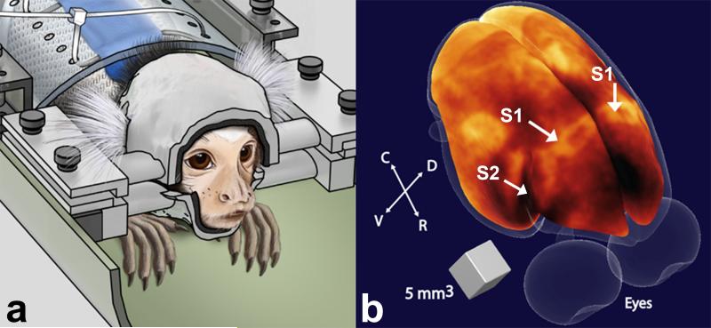
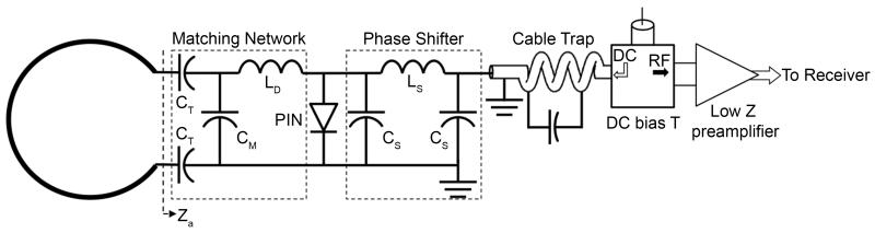
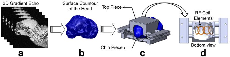
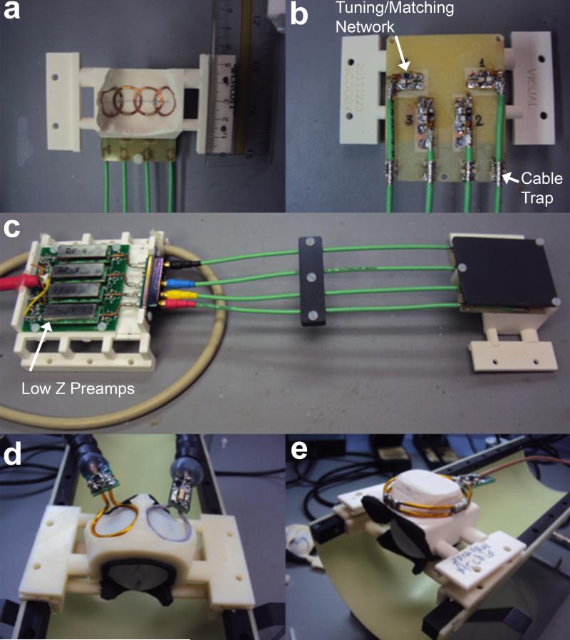
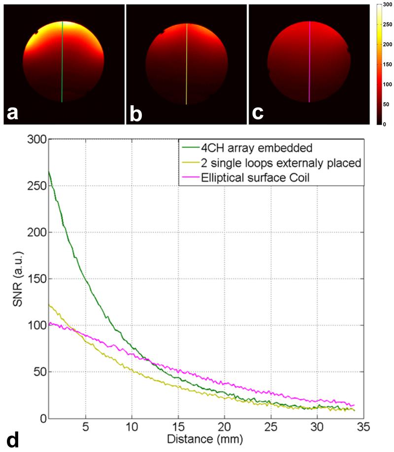
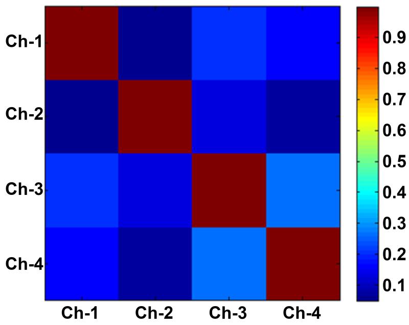
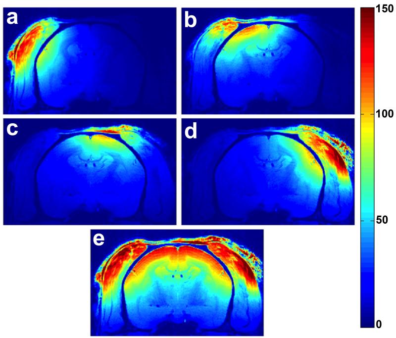
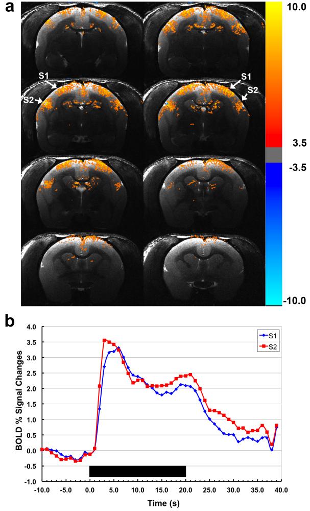
fMRI has established itself as the main research tool in neuroscience and brain cognitive research. The common marmoset (Callithrix jacchus) is a non-human primate model of increasing interest in biomedical research. However, commercial MRI coils for marmosets are not generally available. The present work describes the design and construction of a four-channel receive-only surface RF coil array with excellent signal-to-noise ratio (SNR) specifically optimized for fMRI experiments in awake marmosets in response to somatosensory stimulation. The array was designed as part of a helmet-based head restraint system used to prevent motion during the scans. High SNR was obtained by building the coil array using a thin and flexible substrate glued to the inner surface of the restraint helmet, so as to minimize the distance between the array elements and the somatosensory cortex. Decoupling between coil elements was achieved by partial geometrical overlapping and by connecting them to home-built low-input-impedance preamplifiers. In vivo images show excellent coverage of the brain cortical surface with high sensitivity near the somatosensory cortex. Embedding the coil elements within the restraint helmet allowed fMRI data in response to somatosensory stimulation to be collected with high sensitivity and reproducibility in conscious, awake marmosets.

Keywords: MRI; animal models; marmosets; phased arrays; receive-only RF coils.


Figures

Figure 1
Figure 1
(a) Illustration of a marmoset in the sphinx position restrained by the custom-fit helmet (left). (b) Volumetric rendering of the brain surface of a marmoset obtained from a 3D T1-weighted image (43). Regions of high myelination appear bright in the image. Arrows indicate the locations of primary (S1) and secondary (S2) somatosensory cortex. R = rostral; C = caudal; D = dorsal; V = ventral.
Figure 2
Figure 2
Circuit diagram of one coil element used in the 4-channel embedded array. The electrical length of the RF cable + phase shifter was adjusted to provide 180° phase at the input of the preamplifier. The detuning circuit consisting of CM and LD is activated by providing DC current to the PIN diode via a bias T located before the low input impedance preamplifier.
Figure 3
Figure 3
Schematics of the manufacturing process of the movement restraint helmets. (a) 3D gradient echo images from head and torso are acquired with the body coil in transceiver mode. (b) Surface rendered model obtained from the gradient echo images. (c) From the surface rendered model, the helmets are designed using Rhinoceros 3d and printed in ABS plastic by a 3D printer. (d) 4 overlapped array elements are attached to the inner surface of the helmets, with the matching networks placed on the external side.
Figure 4
Figure 4
(a) Inner surface of helmet showing each element of the embedded 4-channel array. (b) External part of the coil containing the printed circuit board with the matching networks. (c) 4-channel embedded array connected to the low input impedance preamplifiers. For performance evaluation, the embedded 4-channel array was compared to two circular surface coils externally positioned in circular grooves at the helmet top (d), and to a single elliptical surface coil covering the whole marmoset head (e).
Figure 5
Figure 5
Normalized transmit B1+ field maps obtained from a spherical phantom using the linearly driven volume birdcage coil in transceiver mode under two different conditions: (a) In absence of the 4-channel coil array. (b) With the coil array present but with all receive elements actively detuned. The two maps differ from each other by no more than 5%.
Figure 6
Figure 6
Phantom SNR maps calculated from the sum-of-squares images obtained with: (a) the 4-channel embedded array; (b) two single loops externally placed in the helmets; and (c) Elliptical surface coil. (d) Plot of the SNR profiles along the central axis comparing the coils performance.
Figure 7
Figure 7
Noise correlation matrix acquired with the 4-element coil array. The highest off-diagonal correlation coefficient is 0.27, which indicates a small residual coupling between channel 3 and channel 4.
Figure 8
Figure 8
(a-d) In vivo SNR maps obtained for each individual coil element from an awake marmoset. (e) Sum-of-squares image reconstruction showing excellent coil sensitivity in the cortex.
Figure 9
Figure 9
(a) BOLD activation maps obtained from a conscious awake marmoset in response to somatosensory stimulation using the embedded 4-channel array coil. The arrows show the two main regions of activation in the somatosensory cortex, S1 and S2. (b) Normalized averaged time courses showing BOLD signal changes in S1 and S2 (blue and red, respectively). Duration of the stimulation period is indicated by the black bar.

References

    1. Poldrack RA. The role of fMRI in cognitive neuroscience: where do we stand? Curr Opin Neurobiol. 2008;18(2):223–227. - PubMed
    1. Dolan RJ. Neuroimaging of cognition: past, present, and future. Neuron. 2008;60(3):496–502. - PMC - PubMed
    1. Capitanio JP, Emborg ME. Contributions of non-human primates to neuroscience research. Lancet. 2008;371(9618):1126–1135. - PubMed
    1. Mansfield K. Marmoset models commonly used in biomedical research. Comparative Med. 2003;53(4):383–392. - PubMed
    1. Masamoto K, Kim T, Fukuda M, Wang P, Kim SG. Relationship between neural, vascular, and BOLD signals in isoflurane-anesthetized rat somatosensory cortex. Cereb Cortex. 2007;17(4):942–950. - PubMed

Publication types

LinkOut - more resources