. 2013:786:63-78.
doi: 10.1007/978-94-007-6621-1_5.

Stem cells in the Drosophila digestive system

Affiliations

Stem cells in the Drosophila digestive system

Xiankun Zeng et al. Adv Exp Med Biol. 2013.

Abstract

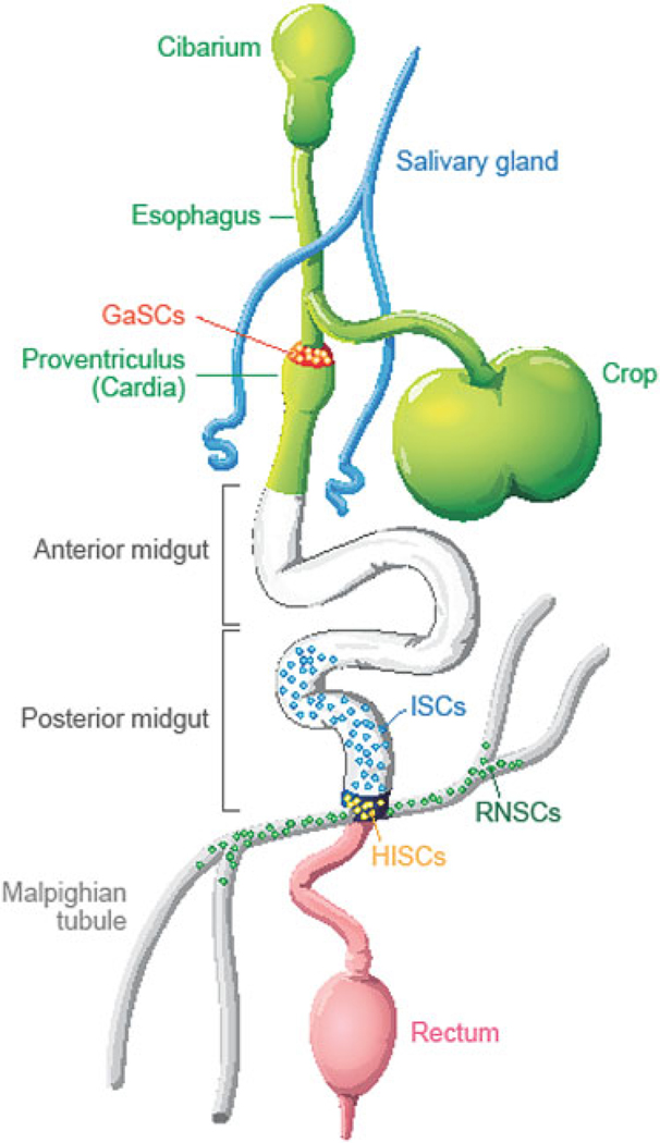
Adult stem cells maintain tissue homeostasis by continuously replenishing damaged, aged and dead cells in any organism. Five types of region and organ-specific multipotent adult stem cells have been identified in the Drosophila digestive system: intestinal stem cells (ISCs) in the posterior midgut; hindgut intestinal stem cells (HISCs) at the midgut/hindgut junction; renal and nephric stem cells (RNSCs) in the Malpighian Tubules; type I gastric stem cells (GaSCs) at foregut/midgut junction; and type II gastric stem cells (GSSCs) at the middle of the midgut. Despite the fact that each type of stem cell is unique to a particular organ, they share common molecular markers and some regulatory signaling pathways. Due to the simpler tissue structure, ease of performing genetic analysis, and availability of abundant mutants, Drosophila serves as an elegant and powerful model system to study complex stem cell biology. The recent discoveries, particularly in the Drosophila ISC system, have greatly advanced our understanding of stem cell self-renewal, differentiation, and the role of stem cells play in tissue homeostasis/regeneration and adaptive tissue growth.


Figures

Fig. 5.1
Fig. 5.1. AMP proliferation and differentiation during Drosophila development.
(a) The adult midgut progenitors (AMP) divide symmetrically to increase their number and remain dispersed as individual cells throughout the midgut during the first two instars. Then each dispersed AMP further divides symmetrically for several rounds to form AMP clusters at the third instar stage. At 3 h after puparium formation (APF), the AMP clusters start fusing together and some of AMPs differentiate into EC. As metamorphosis continues, most of AMPs differentiate into EC and only a few of AMPs remain. These undifferentiated AMPs further divide symmetrically to increase their number and develop into pupal and adult ISC. (b) The Su(H)GBE-Gal4,UAS-mCD8-GFP(green) labeled peripheral cell (PC) extend their process to wrap around the AMP clusters to regulate their proliferation and repress their differentiation. Dl (cytoplasmic red) and Pros (nuclear red) label the AMP and EE respectively
Fig. 5.2
Fig. 5.2. N signaling in ISC regulation.
Dl, specifically expressed in ISC, activates N signaling (N on) at the neighboring EB. Different level of N activity controls the ISC self-renewal and EC versus EE fates. N signaling also controls ISC proliferation
Fig. 5.3
Fig. 5.3. Comparison of asymmetric divisions between SOP and ISC.
Models of asymmetric divisions of SOP and ISC. See text for detail
Fig. 5.4
Fig. 5.4. External signals regulate ISC proliferation and differentiation.
In response to tissue stress and damage JNK, InR, JAK-STAT, EGFR and Wg signaling pathways modulate ISC proliferative response. JNK, InR and JAK-STAT signaling pathways also interact with N signaling to regulate ISC self-renewal and differentiation
Fig. 5.5
Fig. 5.5. Stem cells in adult Drosophila digestive system.
Schematic diagram of Drosophila digestive system including cardia, anterior midgut, posterior midgut, malpighian tubules, hindgut and rectum. Five types of region and organ-specific multipotent adult stem cells have been identified in the Drosophila digestive system: intestinal stem cells (ISCs) in the posterior midgut; hindgut intestinal stem cells (HISCs) at the midgut/hindgut junction; renal and nephric stem cells (RNSCs) in the Malpighian Tubules; type I gastric stem cells (GaSCs) at foregut/midgut junction; and type II gastric stem cells (GSSCs) at the middle of the midgut
Fig. 5.6
Fig. 5.6. Ras-induced stem cell tumor.
(a) GFP labeled RNSC lineage Malpighian tubules. (b) Expression of Rasv12 in RNSC leads to stem cell tumors in Malpighian tubules

References

    1. Weissman IL (2000) Stem cells: units of development, units of regeneration, and units in evolution. Cell 100(1):157–168 - PubMed
    1. Hakim RS, Baldwin K, Smagghe G (2010) Regulation of midgut growth, development, and metamorphosis. Annu Rev Entomol 55:593–608 - PubMed
    1. Saric A, Kalafatic M, Rusak G, Kovacevic G et al. (2007) Postembryonic development of Drosophila melanogaster Meigen, 1830 under the influence of quercetin. Entomol News 118(3):235–240
    1. Slama L, Farkas R (2005) Heartbeat patterns during the postembryonic development of Drosophila melanogaster. J Insect Physiol 51(5):489–503 - PubMed
    1. Yamashita Y (2009) Asymmetric stem cell division and pathology: insights from Drosophila stem cell systems. J Pathol 217(2):181–185 - PubMed

MeSH terms

LinkOut - more resources