. 2013 Aug 15;521(12):2645-Spc1.
doi: 10.1002/cne.23339.

Diverse neuronal lineages make stereotyped contributions to the Drosophila locomotor control center, the central complex

Affiliations

Diverse neuronal lineages make stereotyped contributions to the Drosophila locomotor control center, the central complex

Jacob S Yang et al. J Comp Neurol. .

Abstract

The Drosophila central brain develops from a fixed number of neuroblasts. Each neuroblast makes a clone of neurons that exhibit common trajectories. Here we identified 15 distinct clones that carry larval-born neurons innervating the Drosophila central complex (CX), which consists of four midline structures including the protocerebral bridge (PB), fan-shaped body (FB), ellipsoid body (EB), and noduli (NO). Clonal analysis revealed that the small-field CX neurons, which establish intricate projections across different CX substructures, exist in four isomorphic groups that respectively derive from four complex posterior asense-negative lineages. In terms of the region-characteristic large-field CX neurons, we found that two lineages make PB neurons, 10 lineages produce FB neurons, three lineages generate EB neurons, and two lineages yield NO neurons. The diverse FB developmental origins reflect the discrete input pathways for different FB subcompartments. Clonal analysis enlightens both development and anatomy of the insect locomotor control center.


Figures

Figure 1
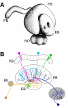
Figure 1. General structures of the central complex (CX) and CX neurons
(A) Sagittal view of the central complex composed of four neuropils: the protocerebral bridge (PB), fan-shape body (FB), ellipsoid body (EB), and noduli (NO) (after Hanesch et al 1989). (B) Schematic illustration of CX small-field and large-field neurons. Line drawing shows a dorsal view of the CX. Three types of small-field neurons, pb-fb-no, fb pontin, and pb-eb neurons, are shown in magenta, cyan, and green, respectively. Two types of large-field neurons, EB ring neuron and LAL (lateral accessory lobe)-fb neuron, are shown in orange and blue, respectively.
Figure 2
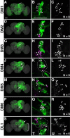
Figure 2. Gross morphologies of DM1–6 and DL1 NB clones
Composite confocal images of representative DM1 (A–C), DM2 (D–F), DM3 (G–I), DM4 (J–L), DM5 (M–O), DM6 (P–R) and DL1 (S–U) NB clones: flip-out MARCM clones shown in an nc82-counterstained standard Drosophila brain (left images), cell body regions of twin-spot MARCM clones with NB progenies shown in green and their paired TAP progenies in magenta (middle images), and the cell bodies of the co-existing TAP clones (right images). In all lineages, clusters of cell bodies are located in posterior brain (arrows in left images). The first sets of larval-derived neuronal progenies (arrowheads in middle and right images) exhibit different specific cell counts with N ranging from 6 to 9. Note presence of two clusters of cell bodies (outlined with dashed lines) in the DM4 and DL1 NB clones, and presence of glia-like progenies in the DL1 NB clone (bracket in S). Clones with cell bodies in right hemispheres were shown in all figures of this study. Scale bars equal 50 μm.
Figure 3
Figure 3. DM1–4 NB clones innervate discrete sets of equivalent CX subdomains
Close-up views of CX elaborations in DM1 (A–C), DM2 (E–G), DM3 (I–K) and DM4 (M–O) NB clones (green), shown at different focal planes from the posterior PB region (left images), through the FB and noduli (middle images), to the anterior EB structure (right images). Their collective patterns of primary CX elaboration are further illustrated in the schematic CXs (D, H, L and P). White arrows: elaboration within the PB; brackets: elaboration within the FB; orange arrows: elaboration within the noduli; solid arrowheads: main elaboration within the EB; open arrowheads: minor elaboration within the EB. Scale bar equals 50 μm.
Figure 4
Figure 4. DM5, DM6 and DL7 NB clones innervate different CX substructures
(A–I) Close-up views of CX elaborations in DM5 (A–C), DM6 (D–F) and DL7 (G–I) NB clones (green), shown at different focal planes from the posterior PB region (left images), through the FB and noduli (middle images), to the anterior EB (right images) structure. The PB elaboration of DM5 is intricately patterned with discrete glomerular concentrations (e.g. arrowheads and bracket in [A]). By contrast, DM6 targets only one PB glomerulus (open arrowhead in [D]), elaborates broadly in layer-specific densities within the FB (bracket in [E]), diffusely innervates the paired noduli (arrowheads in [E]), and occupies the EB with ring-like elaboration (arrow in [F]) plus sector-specific innervation (bracket in [F]). And DL1 selectively targets the top two as well as the bottom two layers of the FB (brackets in [H], corresponding to the 7+6 and the 1+2 layers in [J]). (J) Confocal section of the FB showing layer-specific differential nc82 staining. Note that the alternative weak (w) and strong (S) immunostainings allow division of the FB into seven recognizable layers. (K–L) Confocal sections of the paired noduli at the posterior (K) versus anterior (L) focal plane, revealing three subcompartments: d- dorsal, m- middle, v- ventral. Scale bars equal 50 μm.
Figure 5
Figure 5. Seven additional NB clones with CX elaboration, as revealed by flip-out MARCM
Composite confocal images of representative EBa1 (A–C), LALv1 (D–F), AOTUv4 (G–I), CREa1 (J–L), CREa2 (M–O), SIPp1 (P–R) and PBp1 (S–U) NB clones: flip-out MARCM clones shown in an nc82-counterstained standard Drosophila brain (left images). They innervate various CX substructures, as shown in the confocal sections of EB or PB (middle images) and FB & NO (right images) in the original Z stacks. Various regions of elaboration are pointed with diverse marks. For example, the open arrowhead in [F] indicates the innervation of the contralateral noduli by the LALv1 clone. Scale bars equal 50 μm. n > 3 for all lineages.
Figure 6
Figure 6. Overall projection of seven NB clones with CX elaboration, as revealed by twin-spot MARCM
Composite confocal images of twin-spot MARCM EBa1 (A), LALv1 (B), AOTUv4 (C), CREa1 (D), CREa2 (E), PBp1 (F) and SMPad2 (G) NB clones with NB progenies shown in magenta and their paired GMC progenies in green. The entire clones within the nc82-counterstained brains (blue) are shown. The associated GMC clones contain one or two neurons (yellow arrows) depending on the lineage identity. Scale bar equals 50 μm.
Figure 7
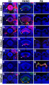
Figure 7. Innervation in CX in the seven additional NB clones with CX elaboration
Innervate various CX substructures, as shown in the confocal sections of EB (left images), FB/NO (middle images) and PB (right images) in the original Z stacks (Figure 6). Innervations of multiple layers of FB are shown with brackets. Note the differential elaborations of the NB clones (while marks) versus their paired GMC clones (yellow marks). Scale bar equals 50 μm.
Figure 8
Figure 8. Neurite trajectories of various NB clones that show CX elaboration (1)
(A–D) Composite confocal images of twin-spot MARCM clones, EBa1 (A–C and M), LALv1 (D–F and N), AOTUv4 (G–I) and CREa1 (J–L), with NB progenies shown in magenta and their paired GMC progenies in green. The entire clones within the nc82-counterstained brains (blue) are shown on the leftmost images. The cell bodies (dashed outlines), primary tracks (directed in arrows with dashed lines that represent out-of-focal-plane projections), and neuropil innervations (indicated with abbreviated neuropil names) are followed from anterior to posterior brain regions as shown from left to right images. Composite confocal images of the 1st larval-born GMC clones (green) of the EBa1 (M) and LALv1 (N) lineages are shown. Note the inneration of multiple neuropils by various single neurons. Scale bar equals 50 μm.
Figure 9
Figure 9. Neurite trajectories of various NB clones that show CX elaboration (2)
(A–C) Composite confocal images of twin-spot MARCM clones, CREa2 (A–C), SMPad2 (D–F) and SIPp1 (G–I), with NB progenies shown in magenta and their paired GMC progenies in green. Composite confocal images of the 1st larval-born GMC clones (green) of the CREa2 (J) and SMPad2 (K) lineages are shown. Scale bar equals 50 μm.
Figure 10
Figure 10. Schematic diagram of possible CX neural projections
Possible wiring patterns within the central complex (small-field neurons, left) and between the CX and other brain neuropils (large-field neurons, right). Lineage origins are shown; dashed lines indicate potential projections.

References

    1. Bayraktar OA, Boone JQ, Drummond ML, Doe CQ. Drosophila type II neuroblast lineages keep Prospero levels low to generate large clones that contribute to the adult brain central complex. Neural Dev. 2010;5:26. - PMC - PubMed
    1. Bello BC, Izergina N, Caussinus E, Reichert H. Amplification of neural stem cell proliferation by intermediate progenitor cells in Drosophila brain development. Neural Dev. 2008;3:5. - PMC - PubMed
    1. Booker R, Truman JW. Postembryonic neurogenesis in the CNS of the tobacco hornworm, Manduca sexta. II. Hormonal control of imaginal nest cell degeneration and differentiation during metamorphosis. J Neurosci. 1987;7(12):4107–4114. - PMC - PubMed
    1. Boone JQ, Doe CQ. Identification of Drosophila type II neuroblast lineages containing transit amplifying ganglion mother cells. Dev Neurobiol. 2008;68(9):1185–1195. - PMC - PubMed
    1. Bowman SK, Rolland V, Betschinger J, Kinsey KA, Emery G, Knoblich JA. The tumor suppressors Brat and Numb regulate transit-amplifying neuroblast lineages in Drosophila. Dev Cell. 2008;14(4):535–546. - PMC - PubMed

Publication types

LinkOut - more resources