. 2013 Jul 19;288(29):21254-21265.
doi: 10.1074/jbc.M113.465138. Epub 2013 May 21.

Breaking the carboxyl rule: lysine 96 facilitates reprotonation of the Schiff base in the photocycle of a retinal protein from Exiguobacterium sibiricum

Affiliations

Breaking the carboxyl rule: lysine 96 facilitates reprotonation of the Schiff base in the photocycle of a retinal protein from Exiguobacterium sibiricum

Sergei P Balashov et al. J Biol Chem. .

Abstract

A lysine instead of the usual carboxyl group is in place of the internal proton donor to the retinal Schiff base in the light-driven proton pump of Exiguobacterium sibiricum (ESR). The involvement of this lysine in proton transfer is indicated by the finding that its substitution with alanine or other residues slows reprotonation of the Schiff base (decay of the M intermediate) by more than 2 orders of magnitude. In these mutants, the rate constant of the M decay linearly decreases with a decrease in proton concentration, as expected if reprotonation is limited by the uptake of a proton from the bulk. In wild type ESR, M decay is biphasic, and the rate constants are nearly pH-independent between pH 6 and 9. Proton uptake occurs after M formation but before M decay, which is especially evident in D2O and at high pH. Proton uptake is biphasic; the amplitude of the fast phase decreases with a pKa of 8.5 ± 0.3, which reflects the pKa of the donor during proton uptake. Similarly, the fraction of the faster component of M decay decreases and the slower one increases, with a pKa of 8.1 ± 0.2. The data therefore suggest that the reprotonation of the Schiff base in ESR is preceded by transient protonation of an initially unprotonated donor, which is probably the ε-amino group of Lys-96 or a water molecule in its vicinity, and it facilitates proton delivery from the bulk to the reaction center of the protein.

Keywords: Bioenergetics; Internal Proton Donor; Photobiology; Proton Pumps; Proton Transport; Retinal Proteins; Spectroscopy.


Figures

FIGURE 1.
FIGURE 1.
Comparison of photocycle transient absorption changes at 410 nm (A), 510 nm (B), and 590 nm (C), in wild type ESR (dotted line) and K96A (solid line) at pH 7.4. The time constants shown on top of A are for the K96A mutant. Samples are in 0.05% lyso-PG, 0.1 m NaCl, 2 mm MOPS.
FIGURE 2.
FIGURE 2.
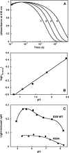
pH dependence of the photocycle and proton transport in the K96A mutant. A, traces 1–4, absorption changes at 410 nm at pH 6.8, 7.4, 7.9, and 8.5, respectively. B, linear dependence of the log of the time constant of M decay versus pH. The slope is 0.63. 0.05% lyso-PG, 0.1 m NaCl, 2 mm MOPS, and 3 mm HEPES. C, light-induced pH changes in suspensions of E. coli cells with wild type ESR and the K96A mutant. The amount of the mutant expressed was ∼1.5-fold larger than that of the wild type. The data for the wild type ESR are from Ref. .
FIGURE 3.
FIGURE 3.
Effect of sodium azide on the kinetics of light-induced absorbance changes in K96A mutant at 410 nm (A) and 590 nm (B). Trace 1, initial sample, pH 7; traces 2–7, upon addition of 20, 50, 100, 200, 400, and 800 mm sodium azide. C, time constant of M decay as a function of sodium azide concentration in K96A and the wild type. 0.05% lyso-PG, 0.5 m NaCl, 10 mm MOPS.
FIGURE 4.
FIGURE 4.
pH dependence of the formation and decay of the M intermediate in wild type ESR. A, traces 1–7, light-induced absorption changes at 410 nm from formation and decay of the M intermediate at pH 6.2, 6.5, 6.8, 7.1, 8.1, 8.4, and 8.7. Traces are normalized at 50 μs. B, traces 1–4, kinetics of M rise and decay in wild type ESR at pH 8.7, 9.2, 10, and 10.3, respectively. C, pH dependence of kinetic components in the decay of the M intermediate. Curves 1 and 2, fast and slower component, respectively. Curve 3, very slow component that appears only above pH 9. D, fractions of the following curves: curve 1, fast (0.8–1 ms); curve 2, slow (8–15 ms), and curve 3, very slow (60 ms and longer) components of M decay.
FIGURE 5.
FIGURE 5.
A, light-induced absorption changes in wild type ESR at 410, 510, 550, and 590 nm at pH 7.1. B and C, difference spectra of the kinetic components of light-induced absorption changes of wild type ESR in lyso-PG at pH 7.1 (B) and 9.3 (C), obtained from global fit of the data with five exponentials. B, spectrum 1, 0.8-ms component corresponding mostly to the decay of M to N1; spectrum 2, 10-ms component from the decay of N1 to N2 plus a minor fraction of M to N1 transition; spectrum 3, 58-ms component from the decay of the N2 to the initial ESR. C, spectrum 1, 7.4-ms component from the decay of M to mostly N2; spectrum 2, 66-ms component from the decay of N2. The fast, 1-ms component of M decay is missing at this pH.
FIGURE 6.
FIGURE 6.
Kinetics of light-induced proton uptake and release in wild type ESR in H2O and D2O; comparison with kinetics of M decay and N rise. A, trace 1, pyranine at pH 7.2 (absorption increase is from proton uptake); trace 2, M intermediate followed at 410 nm; trace 3, formation and decay of the red-shifted intermediates (N1 and N2) at 590 nm (traces normalized). B, same as A but in D2O, pD 7.6. C, effects of replacement of H2O (pH 7.2) with D2O (pD 7.6) on the kinetics of absorption changes at 410 nm from the M intermediate (traces 1 and 2, respectively) and on the kinetics of pyranine absorption changes (traces 3 and 4, respectively). The kinetic components are shown in the Table 2. Traces in D2O are shown as dotted lines. D, kinetics of proton uptake and release at pH 7.2, 8.0, and 9.1, and the kinetics of the Schiff base reprotonation followed at 590 nm in H2O: trace 1, 460 nm, pyranine at pH 7.2; trace 2, 560 nm, phenol red at pH 8.0; trace 3, 595 nm, thymol blue at pH 9.1; trace 4, 590 nm at pH 9.1 in the absence of thymol blue. Traces are normalized. Actual amplitudes were 2.6, 3.6, 3.5, and 26 mOD for the traces 1–4, respectively. The fit of the trace 3 with three components yielded the following time constants: τ1uptake = 650 μs, 23% for fast proton uptake; τ2uptake = 18 ms, 77% for the second phase of uptake, and τrelease = 45 ms for proton release.
FIGURE 7.
FIGURE 7.
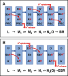
Comparison of the sequences of proton transfer steps (shown with red arrows) in the photocycles of BR (A) and ESR (B). The initial light reactions BR → K, ESR → K, and the transition of K to L are omitted. In both proteins, proton transport involves deprotonation and reprotonation of the retinal Schiff base (Sb) a proton acceptor (A), and a proton donor (D). In BR, additionally, a proton release complex (PRC) has been identified. In BR, A is Asp-85 and D is Asp-96. In ESR, these groups are Asp-85 and Lys-96 or associated water molecule(s). In BR, the M1 to M2 transition involves proton release from the PRC to the bulk, but in ESR, this step involves proton uptake and protonation of D. Therefore, in BR proton release occurs before proton uptake, but in ESR uptake is before release. The key difference between the two proteins is in the way the Schiff base is reprotonated. In BR it receives a proton from the internal donor, which is initially protonated, and reprotonation of the donor occurs during the N1 to N2 transition. In ESR the donor is initially unprotonated, and becomes transiently protonated in the M1 to M2 transition, before it can donate a proton to the Schiff base in the M2 to N1 transition.

References

    1. Petrovskaya L. E., Lukashev E. P., Chupin V. V., Sychev S. V., Lyukmanova E. N., Kryukova E. A., Ziganshin R. H., Spirina E. V., Rivkina E. M., Khatypov R. A., Erokhina L. G., Gilichinsky D. A., Shuvalov V. A., Kirpichnikov M. P. (2010) Predicted bacteriorhodopsin from Exiguobacterium sibiricum is a functional proton pump. FEBS Lett. 584, 4193–4196 - PubMed
    1. Balashov S. P., Petrovskaya L. E., Lukashev E. P., Imasheva E. S., Dioumaev A. K., Wang J. M., Sychev S. V., Dolgikh D. A., Rubin A. B., Kirpichnikov M. P., Lanyi J. K. (2012) Aspartate-histidine interaction in the retinal Schiff base counterion of the light-driven proton pump of Exiguobacterium sibiricum. Biochemistry 51, 5748–5762 - PMC - PubMed
    1. Vishnivetskaya T., Kathariou S., McGrath J., Gilichinsky D., Tiedje J. M. (2000) Low-temperature recovery strategies for the isolation of bacteria from ancient permafrost sediments. Extremophiles 4, 165–173 - PubMed
    1. Rodrigues D. F., Goris J., Vishnivetskaya T., Gilichinsky D., Thomashow M. F., Tiedje J. M. (2006) Characterization of Exiguobacterium isolates from the Siberian permafrost. Description of Exiguobacterium sibiricum sp. nov. Extremophiles 10, 285–294 - PubMed
    1. Oesterhelt D., Stoeckenius W. (1973) Functions of a new photoreceptor membrane. Proc. Natl. Acad. Sci. U.S.A. 70, 2853–2857 - PMC - PubMed

Publication types

MeSH terms

LinkOut - more resources