. 2013 May 28;110(22):8948-53.
doi: 10.1073/pnas.1220637110. Epub 2013 May 21.

Brief demethylation step allows the conversion of adult human skin fibroblasts into insulin-secreting cells

Affiliations

Brief demethylation step allows the conversion of adult human skin fibroblasts into insulin-secreting cells

Georgia Pennarossa et al. Proc Natl Acad Sci U S A. .

Abstract

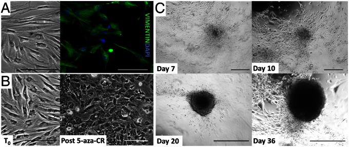
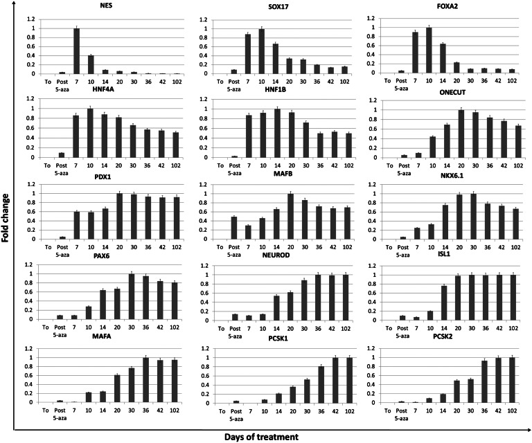
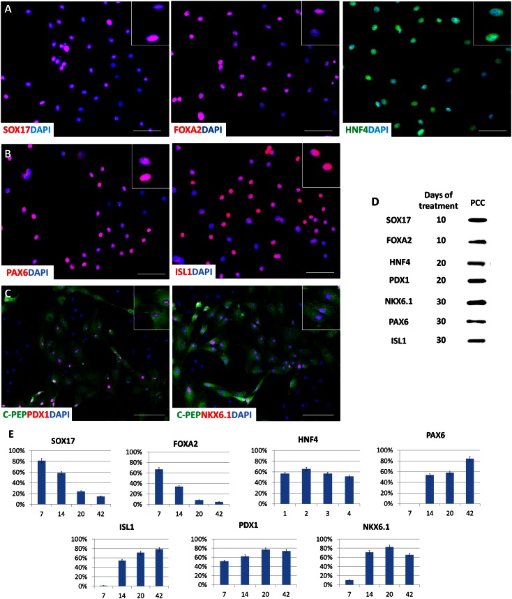
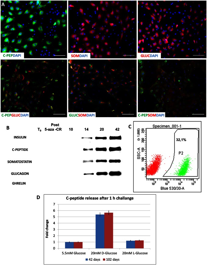
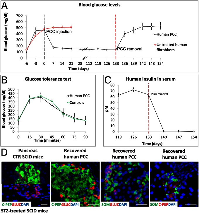
The differentiated state of mature cells of adult organisms is achieved and maintained through the epigenetic regulation of gene expression, which consists of several mechanisms including DNA methylation. The advent of induced pluripotent stem cell technology enabled the conversion of adult cells into any other cell type passing through a stable pluripotency state. However, indefinite pluripotency is unphysiological, inherently labile, and makes cells prone to culture-induced alterations. The direct conversion of one cell type to another without an intermediate pluripotent stage is also possible but, at present, requires the viral transfection of appropriate transcription factors, limiting its therapeutic potential. The aim of this study was to investigate whether it is possible to achieve the direct conversion of an adult cell by exposing it to a demethylating agent immediately followed by differentiating culture conditions. Adult human skin fibroblasts were exposed for 18 h to the DNA methyltransferase inhibitor 5-azacytidine, followed by a three-step protocol for the induction of endocrine pancreatic differentiation that lasted 36 d. At the end of this treatment, 35 ± 8.9% fibroblasts became pancreatic converted cells that acquired an epithelial morphology, produced insulin, and then released the hormone in response to a physiological glucose challenge in vitro. Furthermore, pancreatic converted cells were able to protect recipient mice against streptozotocin-induced diabetes, restoring a physiological response to glucose tolerance tests. This work shows that it is possible to convert adult fibroblasts into insulin-secreting cells, avoiding both a stable pluripotent stage and any transgenic modification.

Keywords: cell plasticity; pancreatic beta cell.


Conflict of interest statement

The authors declare no conflict of interest.

Figures

Fig. 1.
Fig. 1.
Morphological changes in adult human skin fibroblasts exposed to 5-aza-CR and subjected to endocrine pancreatic induction. (A) Immunolocalization of vimentin, the typical fibroblast intermediate filament protein. Uniform positivity indicated a homogenous cell population at the onset of the experiments. (Scale bar, 200 µm.) (B) Untreated cells (T0) underwent marked morphological changes in response to an 18-h exposure to 5-aza-CR (post 5-aza-CR). Fibroblasts changed their typical elongated shape into a round epithelioid aspect. Cell size was smaller, and nuclei became larger and more granular. (Scale bar, 200 µm.) (C) Representative pictures of the morphological changes taking place during endocrine pancreatic induction. Cells exposed to activin A gradually organized in clusters (day 7). In response to the addition of retinoic acid, they rearranged in a reticular pattern and clustered in distinguishable aggregates (day 10). These formations progressed with time and were further stimulated by B27/bFGF/ITS, which led to the recruitment of a growing number of cells, aggregating in large 3D colonies (day 20). Finally, colonies became spherical structures that tended to detach and float freely in the culture medium, reminiscent of typical pancreatic islets in vitro (day 36). (Scale bar, 400 µm.)
Fig. 2.
Fig. 2.
Gene expression changes in adult human skin fibroblasts exposed to 5-aza-CR and subjected to endocrine pancreatic induction. Expression pattern of markers of early (NES, SOX17, FOXA2, HNF4A, HNF1B, ONECUT, PDX1, and MAFB) and mature pancreatic precursors (NKX6.1, PAX6, NEUROD, ISL1, MAFA, PCSK1, and PCSK2) in untreated fibroblasts (T0), in fibroblast exposed to 5-aza-CR (post 5-aza-CR), and at different days of pancreatic induction (days 7–102).
Fig. 3.
Fig. 3.
Immunocytochemical localization of endoderm and pancreatic markers during human skin fibroblasts’ conversion to endocrine pancreatic cells. (A) Immunolocalization of endoderm (SOX17, FOXA2) and primitive gut tube (HNF4) markers in fibroblasts subjected to pancreatic induction for 10 d. (B) Immunolocalization of PAX6 and ISL1, markers of more advanced pancreatic differentiation, on day 30 after exposure to 5-aza-CR. (C) Colocalization of PDX1 and NKX6.1 with C-peptide was also observed on day 30 after exposure to 5-aza-CR. (D) Western blot immunodetection of SOX17, FOXA2, HNF4, PDX1, NKX6.1, PAX6, and ISL1 in PCCs on different days of treatment. (E) Proportion of positive cells for the different molecules during the differentiation process in vitro. (Scale bars, 200 µm.)
Fig. 4.
Fig. 4.
Morphologic characterization of human PCCs. (A) Immunostaining of PCCs after 36 d of culture reveals a clear signal of C-peptide, somatostatin, and glucagon in positive cells (Upper). Although some cells expressing only C-peptide can be seen, most cells are positive for more than one hormone (Lower). (Scale bar, 200 µm.) (B) Representative Western blot analysis of constitutive proteins collected at different times of culture. Insulin, C-peptide, somatostatin, and glucagon were detected from day 14 and steadily increased. Consistent with the absence of its mRNA, ghrelin was not detectable. β-Actin was used to check that an equal protein amount was loaded on each lane. (C) Representative output of flow cytometer analysis showing the efficiency toward β-cell differentiation measured counting C-peptide-labeled cells. (D) Quantification of C-peptide release in the culture medium in response to 20 mM d-glucose for 1 h at a different time of culture. Bars represents the mean ± SD of three independent replicates.
Fig. 5.
Fig. 5.
Functional characterization of human PCCs. (A) S.c. injection of 5 × 10⁶ PCCs in STZ-treated SCID mice promptly decreased their glucose blood levels. Glucose levels remained constant up to 133 d. Injection of the same number of untreated fibroblasts did not elicit any effect, and mice died after 4 wk. Removal of PCCs from STZ-treated mice caused a rapid rise in glycemic values, indicating that PCCs were the functional source of insulin. (B) I.p. injection of 3 g per kilogram body weight induced a rise of blood glucose concentration that returned to basal level within 90 min, both in PCC-engrafted and control mice. The test was repeated 3 times at 1-wk intervals. (C) Levels of human insulin in the serum of STZ-treated mice during PCC engraftment and after its removal. (D) Immunolocalization of C-peptide and glucagon in pancreatic islets of control and STZ SCID mice indicate the selective destruction of beta cells. (Scale bar, 20 µm.) Immunolocalization of C-peptide, glucagon, and somatostatin in human PCCs removed from SCID mice. Merged costaining demonstrates that each cell produces a single hormone. (Scale bar, 50 µm.)

References

    1. Lengner CJ. iPS cell technology in regenerative medicine. Ann N Y Acad Sci. 2010;1192:38–44. - PubMed
    1. Cohen DE, Melton D. Turning straw into gold: Directing cell fate for regenerative medicine. Nat Rev Genet. 2011;12(4):243–252. - PubMed
    1. Kiskinis E, Eggan K. Progress toward the clinical application of patient-specific pluripotent stem cells. J Clin Invest. 2010;120(1):51–59. - PMC - PubMed
    1. Li W, Ding S. Small molecules that modulate embryonic stem cell fate and somatic cell reprogramming. Trends Pharmacol Sci. 2010;31(1):36–45. - PubMed
    1. Ding S, Schultz PG. A role for chemistry in stem cell biology. Nat Biotechnol. 2004;22(7):833–840. - PubMed

Publication types

MeSH terms