. 2013 Jun 4;110(23):9583-8.
doi: 10.1073/pnas.1303483110. Epub 2013 May 21.

Single-membrane-bounded peroxisome division revealed by isolation of dynamin-based machinery

Affiliations

Single-membrane-bounded peroxisome division revealed by isolation of dynamin-based machinery

Yuuta Imoto et al. Proc Natl Acad Sci U S A. .

Abstract

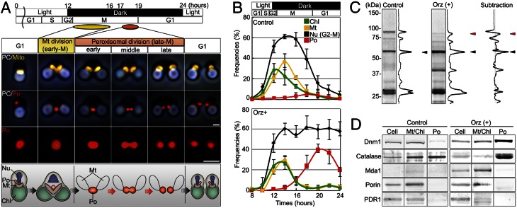
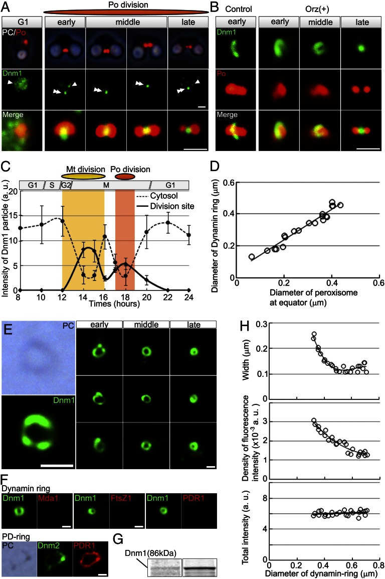
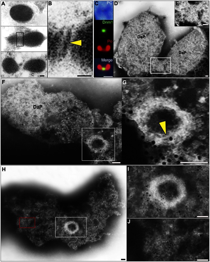
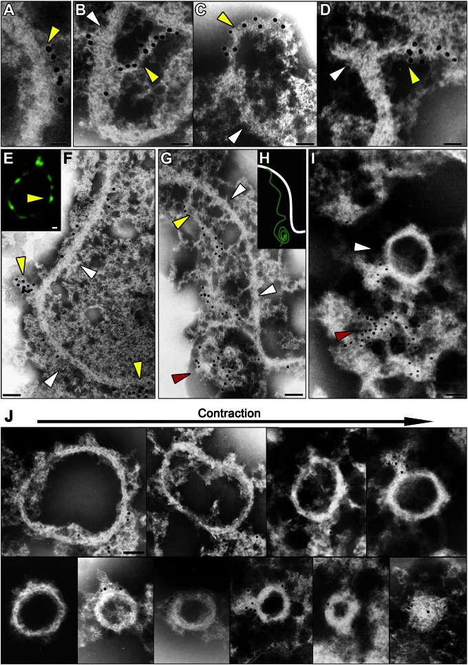
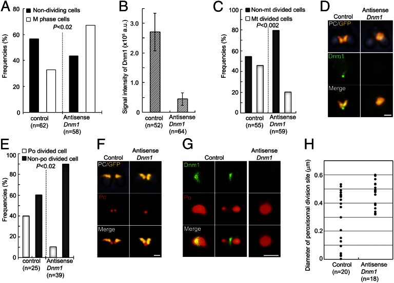
Peroxisomes (microbodies) are ubiquitous single-membrane-bounded organelles and fulfill essential roles in the cellular metabolism. They are found in virtually all eukaryotic cells and basically multiply by division. However, the mechanochemical machinery involved in peroxisome division remains elusive. Here, we first identified the peroxisome-dividing (POD) machinery. We isolated the POD machinery from Cyanidioschyzon merolae, a unicellular red alga containing a single peroxisome. Peroxisomal division in C. merolae can be highly synchronized by light/dark cycles and the microtubule-disrupting agent oryzalin. By proteomic analysis based on the complete genome sequence of C. merolae, we identified a dynamin-related protein 3 (DRP3) ortholog, CmDnm1 (Dnm1), that predominantly accumulated with catalase in the dividing-peroxisome fraction. Immunofluorescence microscopy demonstrated that Dnm1 formed a ring at the division site of the peroxisome. The outlines of the isolated dynamin rings were dimly observed by phase-contrast microscopy and clearly stained for Dnm1. Electron microscopy revealed that the POD machinery was formed at the cytoplasmic side of the equator. Immunoelectron microscopy showed that the POD machinery consisted of an outer dynamin-based ring and an inner filamentous ring. Down-regulation of Dnm1 impaired peroxisomal division. Surprisingly, the same Dnm1 serially controlled peroxisomal division after mitochondrial division. Because genetic deficiencies of Dnm1 orthologs in multiperoxisomal organisms inhibited both mitochondrial and peroxisomal proliferation, it is thought that peroxisomal division by contraction of a dynamin-based machinery is universal among eukaryotes. These findings are useful for understanding the fundamental systems in eukaryotic cells.


Conflict of interest statement

The authors declare no conflict of interest.

Figures

Fig. 1.
Fig. 1.
Identification of Dnm1 from the dividing-peroxisome fraction. (A) Immunofluorescence and schematic images of mitochondrial and peroxisomal divisions of C. merolae. Peroxisomal (red) division occurred after mitochondrial (yellow) division. Chl, chloroplast; Mt, mitochondrion; Nu, nucleus; PC, phase-contrast image; Po, peroxisome. (B) Frequencies of dividing cell nuclei (Nu), dividing chloroplasts (Chl), dividing mitochondria (Mt), and dividing peroxisomes (Po) in non–oryzalin-treated cells (control) and oryzalin-treated cells (Orz+) at the indicated times after synchronization (n > 100). (C) Proteomic analysis of peroxisomal fractions in control and oryzalin-treated cells at 20 h after synchronization. The major bands specific to oryzalin-treated cells were identified as catalase (black arrowhead), Dnm1 (red arrowhead), and others. (D) Immunoblot analyses of Dnm1, catalase, mitochondria division protein (Mda1), porin, and chloroplast division protein (PDR1). Cell, whole cell; Mt/Chl, isolated mitochondria and chloroplast; Po, isolated peroxisomes. (Scale bars: 1 μm.)
Fig. 2.
Fig. 2.
Dynamin localization at the peroxisome division site and isolation of dynamin rings. (A) Phase-contrast and immunofluorescence images of the peroxisome (Po) (red) and Dnm1 (green) and enlarged merged images. (B) Immunofluorescence images of the dynamin ring-like structures (green) encircling dividing peroxisomes (red) in slightly squashed control and oryzalin-treated cells. (C) Dynamic change of Dnm1 signals between the cytosol (dotted line) and division site of mitochondrion or peroxisome (solid line) during mitochondrial and peroxisomal divisions (n > 50). (D) The diameter of the dynamin ring decreases as the diameter of the division site decreases during the peroxisome division process. (E) Phase-contrast and immunofluorescence images of closed dynamin rings (green) isolated from peroxisomes at various stages of division. (F) Immunofluorescence images of isolated dynamin rings from dividing peroxisomal fraction showing Dnm1 (green) and Mda1, FtsZ1, or PDR1 (red). Images of isolated PD ring showing Dnm2 (green) and PDR1 (red) as a control. (G) SDS/PAGE of the dividing-peroxisome fraction (Left) and isolated dynamin ring fraction (Right). (H) Width, density of intensity, and total fluorescence intensity of the Dnm1 signals relative to the diameter of the dynamin ring. White arrowheads, cytosolic dynamin; double arrowhead, division site. [Scale bars: 1 μm (A and B); 500 nm (E and F).]
Fig. 3.
Fig. 3.
Identification of the POD machinery in C. merolae cells by thin-section EM and immuno-EM. (A and B) Thin-section EM images showing the progress of peroxisomal division. The edge of the POD machinery was visualized as a fine bar on the cytoplasmic side of the single membrane (yellow arrowhead) at the peroxisome division site. (C) Immunofluorescence images showing Dnm1 (green) localized at the division plane of isolated peroxisomes (red). (D and E) Immuno-EM images showing a parallel image to the division plane. POD machinery containing Dnm1 was visible on bridges between the daughter peroxisomes. (F and G) Perpendicular image to the division plane. A filamentous ring (yellow arrowhead) appears after dissociation of the membrane. (H and I) Dnm1 is localized on the periphery of the POD machinery, and catalase is localized on the membrane. (J) Enlarged micrograph of the red boxed area in H showing partially dissolved peroxisome membrane labeled with catalase. Detergent treatment for 30 s (CI); DaP, daughter peroxisome; large immunogold particles (15 nm), Dnm1 (CJ); small immunogold particles (10 nm), catalase (HJ). B, E, G, and I show enlarged micrographs of the boxed areas in A, D, F, and H, respectively. [Scale bars: 50 nm (A and B, FH); 1μm (C); 10nm (D and E).]
Fig. 4.
Fig. 4.
Immuno-EM and immunofluorescence microscopic images and schematic representation showing the separation of the filamentous ring and the dynamin-based ring. AD, F, G, I, and J and E and H show immuno-EM and fluorescence microscopic images, respectively. (A) Part of the dynamin-based ring (yellow arrowheads) is arranged at distances of about 10 nm along the filamentous ring (white arrowheads) in the POD machinery. (BD) The dynamin-based rings (yellow arrowheads) are peeled off from the filamentous ring (white arrowheads) in parts of the POD machinery. (E and F) The separated dynamin rings form clumps (yellow arrowheads) on the filamentous ring (white arrowheads). (G) The dynamin-based ring separated from the filamentous ring (white arrowheads) shows a string (yellow arrowheads) and a part of the ring forms a clump (red arrowhead), suggesting that the dynamin-based ring encircles the filamentous ring. (H) The schematic representation shows the dynamin-based ring (green line) and the filamentous ring (white line). (I) The dynamin-based ring separates from the filamentous ring (white arrowhead). (J) Isolated POD machinery arranged in order of division. Immunogold particles (15 nm), Dnm1 (AD, F, and G); green, anti-Dnm1 antibody (E). [Scale bars: 50 nm (AI); 100 nm (J).]
Fig. 5.
Fig. 5.
Down-regulation of Dnm1. (A) Frequencies of nondividing cells (interphase) and M phase cells. Nondividing cells are decreased, whereas M phase cells are increased, at 48 h after introduction of the antisense-Dnm1 DNA (P < 0.02 by Fisher’s exact test). (B) The immunofluorescence intensity of the dynamin ring is decreased by the down-regulation. Data are means and SD (n > 50). (C) Down-regulation of Dnm1 inhibits mitochondrial division (n > 50; P < 0.002 by Fisher’s exact test). (D) Phase-contrast and immunofluorescence images showing a dividing mitochondrion (GFP, yellow, anti-GFP) and Dnm1 (green) in control cells and a nondivided mitochondrion without dynamin in antisense-Dnm1 DNA-treated cells. (E) Cells with down-regulated Dnm1 show inhibited peroxisomal division, and the number of non–peroxisome-dividing cells is increased during late M phase (P < 0.02 by Fisher’s exact test). Data are the frequencies of control and antisense-Dnm1 DNA-treated cells with dividing peroxisomes (po) and nondividing peroxisomes (n > 25). (F) Phase-contrast and immunofluorescence images showing a dividing mitochondrion with a dividing peroxisome in control cells and a dividing mitochondrion with a nondividing peroxisome in antisense-Dnm1 DNA-treated cells. (G) Immunofluorescence images show a dividing peroxisome (red) with a dynamin ring (green) in control cells and a nondividing oval-shaped peroxisome without a dynamin ring in Dnm1-down-regulated cells. (H) Peroxisomal division in Dnm1–down-regulated cells stops at the early stage of contraction of the POD machinery. The data represent the diameters of the peroxisome division sites in control cells and antisense-Dnm1 DNA-treated cells. (Scale bars: 1 μm.)

References

    1. Schrader M, Yoon Y. Mitochondria and peroxisomes: Are the ‘big brother’ and the ‘little sister’ closer than assumed? Bioessays. 2007;29(11):1105–1114. - PubMed
    1. Pan R, Hu J. The conserved fission complex on peroxisomes and mitochondria. Plant Signal Behav. 2011;6(6):870–872. - PMC - PubMed
    1. Gould SJ, Valle D. Peroxisome biogenesis disorders: Genetics and cell biology. Trends Genet. 2000;16(8):340–345. - PubMed
    1. Motley AM, Hettema EH. Yeast peroxisomes multiply by growth and division. J Cell Biol. 2007;178(3):399–410. - PMC - PubMed
    1. Koch A, Schneider G, Lüers GH, Schrader M. Peroxisome elongation and constriction but not fission can occur independently of dynamin-like protein 1. J Cell Sci. 2004;117(Pt 17):3995–4006. - PubMed

Publication types

LinkOut - more resources