

Medial posterior meniscal root tears are associated with development or worsening of medial tibiofemoral cartilage damage: the multicenter osteoarthritis study
- PMID: 23696679
- PMCID: PMC3750419
- DOI: 10.1148/radiol.13122544
Medial posterior meniscal root tears are associated with development or worsening of medial tibiofemoral cartilage damage: the multicenter osteoarthritis study
Abstract
Purpose: To assess the association of meniscal root tear with the development or worsening of tibiofemoral cartilage damage.
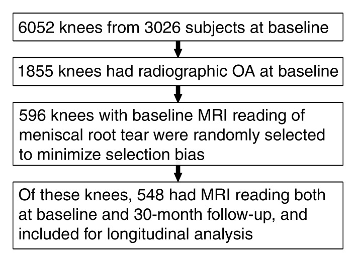
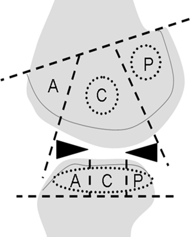
Materials and methods: Institutional review board approval and written informed consent from all subjects were obtained. A total of 596 knees with radiographically depicted osteoarthritis were randomly selected from the Multicenter Osteoarthritis study cohort. Cartilage damage was semiquantitatively assessed by using the Whole-Organ Magnetic Resonance Imaging Score (WORMS) system (grades 0-6). Subjects were separated into three groups: root tear only, meniscal tear without root tear, and neither meniscal nor root tear. A log-binomial regression model was used to calculate the relative risks for knees to develop incident or progressing cartilage damage in the root tear group and the meniscal tear group, with the no tear group serving as a reference.
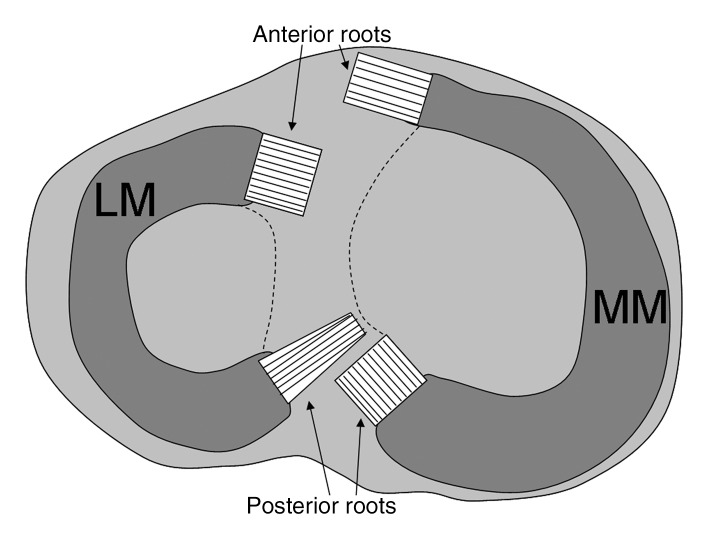
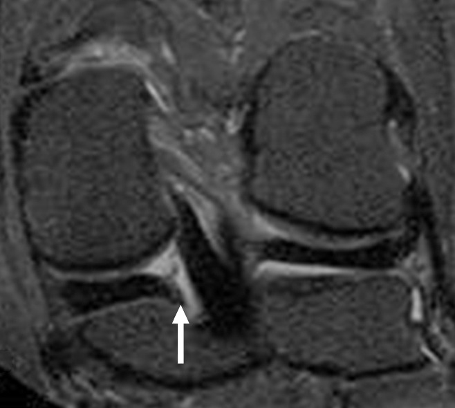
Results: In the medial tibiofemoral joint, there were 37 knees with isolated medial posterior root tear, 294 with meniscal tear without root tear, and 264 without meniscal or root tear. There were only two lateral posterior root tears, and no anterior root tears were found. Thus, the focus was on the medial posterior root tear. The frequency of severe cartilage damage (WORMS ≥ 5) was higher in the group with root tear than in the group without root or meniscal tear (76.7% vs 19.7%, P < .0001) but not in the group with meniscal but no root tear (76.7% vs 65.2%, P = .055). Longitudinal analyses included 33 knees with isolated medial posterior root tear, 270 with meniscal tear, and 245 with no tear. Adjusted relative risk of cartilage loss was 2.03 (95% confidence interval [CI]: 1.18, 3.48) for the root tear group and 1.84 (95% CI: 1.32, 2.58) for the meniscal tear group.
Conclusion: Isolated medial posterior meniscal root tear is associated with incident and progressive medial tibiofemoral cartilage loss.
Figures


References
-
- Soames RW. Skeletal system. In: Williams PL, ed. Gray’s anatomy. 38th ed New York, NY: Churchill Livingstone, 1995; 720–724
-
- Koenig JH, Ranawat AS, Umans HR, Difelice GS. Meniscal root tears: diagnosis and treatment. Arthroscopy 2009;25(9):1025–1032 - PubMed
-
- Pagnani MJ, Cooper DE, Warren RF. Extrusion of the medial meniscus. Arthroscopy 1991;7(3):297–300 - PubMed
-
- Choi CJ, Choi YJ, Lee JJ, Choi CH. Magnetic resonance imaging evidence of meniscal extrusion in medial meniscus posterior root tear. Arthroscopy 2010;26(12):1602–1606 - PubMed
-
- Lerer DB, Umans HR, Hu MX, Jones MH. The role of meniscal root pathology and radial meniscal tear in medial meniscal extrusion. Skeletal Radiol 2004;33(10):569–574 - PubMed
Publication types
MeSH terms
Grants and funding
LinkOut - more resources
Full Text Sources
Other Literature Sources
Medical
