. 2013;9(5):e1003364.
doi: 10.1371/journal.ppat.1003364. Epub 2013 May 16.

Structural basis of HCV neutralization by human monoclonal antibodies resistant to viral neutralization escape

Affiliations

Structural basis of HCV neutralization by human monoclonal antibodies resistant to viral neutralization escape

Thomas Krey et al. PLoS Pathog. 2013.

Abstract

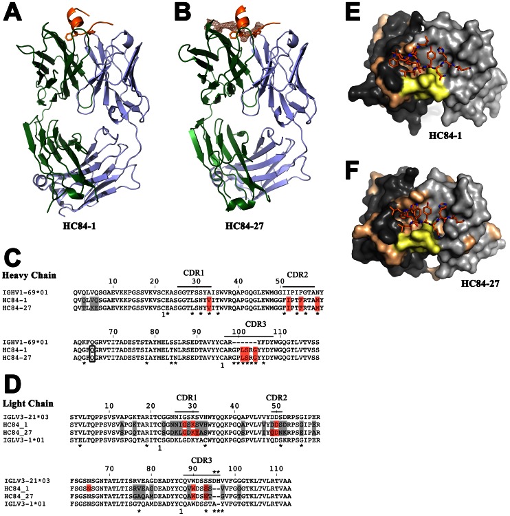
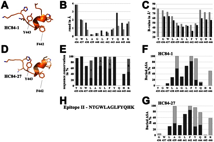
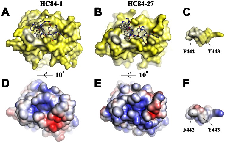
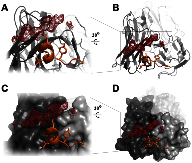
The high mutation rate of hepatitis C virus allows it to rapidly evade the humoral immune response. However, certain epitopes in the envelope glycoproteins cannot vary without compromising virus viability. Antibodies targeting these epitopes are resistant to viral escape from neutralization and understanding their binding-mode is important for vaccine design. Human monoclonal antibodies HC84-1 and HC84-27 target conformational epitopes overlapping the CD81 receptor-binding site, formed by segments aa434-446 and aa610-619 within the major HCV glycoprotein E2. No neutralization escape was yet observed for these antibodies. We report here the crystal structures of their Fab fragments in complex with a synthetic peptide comprising aa434-446. The structures show that the peptide adopts an α-helical conformation with the main contact residues F⁴⁴² and Y⁴⁴³ forming a hydrophobic protrusion. The peptide retained its conformation in both complexes, independently of crystal packing, indicating that it reflects a surface feature of the folded glycoprotein that is exposed similarly on the virion. The same residues of E2 are also involved in interaction with CD81, suggesting that the cellular receptor binds the same surface feature and potential escape mutants critically compromise receptor binding. In summary, our results identify a critical structural motif at the E2 surface, which is essential for virus propagation and therefore represents an ideal candidate for structure-based immunogen design for vaccine development.


Conflict of interest statement

We have read the journal's policy and have the following conflicts. FAR received recurrent funding from Merck-Serono. The funders did not participate in the design and conduct of the study; collection, analysis, and interpretation of the data; and preparation, review, or approval of the manuscript. This funding does not alter our adherence to all PLOS Pathogens policies on sharing data and materials. The other authors declare that they have no conflict of interest.

Figures

Figure 1
Figure 1. Crystal structure of HC84 mAbs in complex with their peptide epitope.
Structure of mAbs HC84-1 (A) and HC84-27 (B) in complex with a peptide mimicking the epitope II of HCV strain H77. The crystal structures of two broadly neutralizing antibodies – HC84-1 (crystallized in space-group C2221) and HC84-27 (crystallized in spacegroup P1) in complex with its epitope were determined and refined to 2.1 Å resolution and 2.2 Å resolution, respectively. The crystal structures are shown as cartoon. The two aromatic residues F442 and Y443 (side chains shown as sticks) at the C-terminal end of the α-helix formed by the peptide (orange) insert into a cavity formed by the heavy chain (green) CDR loops. The peptide C-terminal extended region (Y443-K446) contacts the CDR loops of the light chain (blue). Electron density of the Fo-Fc map of the HC84-27 complex is contoured at 2 σ around the second binding site, indicating its relative position to epitope II. (C+D) Amino acid sequences of the variable region of the heavy (C) and light (D) chain of the two mAbs and the respective closest germ-line homologue suggested by IMGT V-QUEST and junction analysis were aligned. The disulfide connectivity is numbered and indicated below the alignment. Bars show the positions of the CDRs according to the IMGT nomenclature . Asterisks below the sequence indicate the position of somatic mutations and the junction region in the CDR-H3 loop, respectively. Amino acid differences between the two mAbs are shaded in light grey and boxed residues represent contact residues with the epitope II peptide (red). (E+F) View on the paratopes of Fab HC84-1 (E) and HC84-27 (F), respectively. The molecular surface of the light chain and heavy chain are colored in light grey and dark grey, respectively. Somatic mutations and the junction region in CDR-H3 resulting from V-D-J recombination are mapped on the surface and colored in beige and yellow, respectively.
Figure 2
Figure 2. Analysis of epitope II peptide structure.
Crystal structure of the epitope II peptide from the complex with HC84-1 (A) and the complex with HC84-27 (D) is shown as cartoon displaying the side chains as sticks. View on the side of the peptide facing the paratope. Residues W437-F442 make a 1.5 α-helical turn followed by an extended segment containing residues Y443-K446. (B) Root mean square deviation (rmsd) upon superposition of the peptides using Chimera including all atoms (dark grey) or main chain atoms only (light grey) in the calculation and represented per residue (C) Average temperature factors of the peptides plotted per residue for complexes with HC84-1 (light grey) and HC84-27 (dark grey). (E) HCV sequence conservation calculated across 6,998 isolates for this region and analyzed with the ViPR database (http://www.viprbrc.org). For residues L438, G440, F442 and K446, alternative abundant residues are shown as stacked columns in grey and light grey. (F+G) Percentages of accessible surface area (ASA) buried in the complex, calculated using PISA represented per residue as stacked columns for heavy (dark grey) and light (light grey) chains of the respective Fabs. (H) Amino acid sequence of the epitope II peptide from strain H77.
Figure 3
Figure 3. Interaction between HC84 Fabs and the epitope II peptide.
The paratopes on Fab HC84-1 (A+D) and HC84-27 (B+E) are shown as molecular surface. (A+B) The epitope II peptide shown as sticks and colored by atom-type (grey, red and blue for carbon, oxygen and nitrogen, respectively). (A−C) The molecular surface of the paratope (A−B) and the epitope peptide (C) colored according to a normalized hydrophobicity scale from white (hydrophobic) to bright yellow (hydrophilic). (D−F) Electrostatic potential [−5 kT/e (red) to 5 kT/e (blue)] across the molecular surfaces of the paratopes (D−E) and the peptide epitope (F) calculated using the adaptive Poisson-Boltzmann solver. (C+F) The molecular surface of the peptide is shown looking from the paratope. The hydrophobic protrusion constituted by F442 and Y443 inserts into the cavity formed by the CDR H1-3 loops and framework residues around the CDR-H2 loop.
Figure 4
Figure 4. Position of the second antigen binding site on HC84-27.
View on the paratope of HC84-27 in complex with the epitope II peptide in overview (B+D) and in detail (A+C). The light chain and heavy chain are colored in light grey and dark grey, respectively. Fab HC84-27 is shown in cartoon representation (A+B) or as molecular surface (C+D). The epitope II peptide is shown as cartoon with F442 and Y443 side chains shown as sticks and colored by atom type (as in Figure 2). Electron density of the Fo-Fc map is contoured at 2 σ, indicating the position of the second antigen binding site close to β-strand C″, which is labeled.

References

    1. Lemon SM, Walker CM, Alter MJ, Yi MK (2007) Hepatitis C Virus. Fields Virology, Fifth edition 1253–1304.
    1. Verna EC, Brown RS Jr (2008) Hepatitis C and liver transplantation: enhancing outcomes and should patients be retransplanted. Clin Liver Dis 12: 637–659, ix–x. - PubMed
    1. Simmonds P, Bukh J, Combet C, Deléage G, Enomoto N, et al. (2005) Consensus proposals for a unified system of nomenclature of hepatitis C virus genotypes. Hepatology (Baltimore, Md) 42: 962–973. - PubMed
    1. Kwo PY, Lawitz EJ, McCone J, Schiff ER, Vierling JM, et al. (2010) Efficacy of boceprevir, an NS3 protease inhibitor, in combination with peginterferon alfa-2b and ribavirin in treatment-naive patients with genotype 1 hepatitis C infection (SPRINT-1): an open-label, randomised, multicentre phase 2 trial. The Lancet 376: 705–716. - PubMed
    1. McHutchison JG, Manns MP, Muir AJ, Terrault NA, Jacobson IM, et al. (2010) Telaprevir for previously treated chronic HCV infection. New England Journal of Medicine 362: 1292–1303. - PubMed

Publication types

MeSH terms