. 2013;9(5):e1003374.
doi: 10.1371/journal.ppat.1003374. Epub 2013 May 16.

Orthobunyavirus ultrastructure and the curious tripodal glycoprotein spike

Affiliations

Orthobunyavirus ultrastructure and the curious tripodal glycoprotein spike

Thomas A Bowden et al. PLoS Pathog. 2013.

Abstract

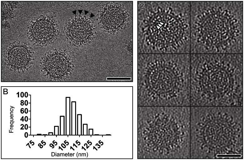
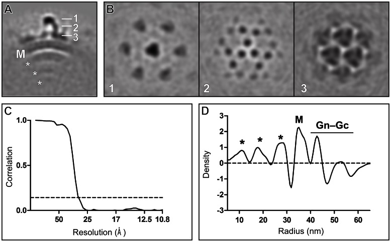
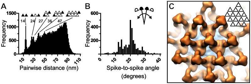
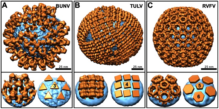
The genus Orthobunyavirus within the family Bunyaviridae constitutes an expanding group of emerging viruses, which threaten human and animal health. Despite the medical importance, little is known about orthobunyavirus structure, a prerequisite for understanding virus assembly and entry. Here, using electron cryo-tomography, we report the ultrastructure of Bunyamwera virus, the prototypic member of this genus. Whilst Bunyamwera virions are pleomorphic in shape, they display a locally ordered lattice of glycoprotein spikes. Each spike protrudes 18 nm from the viral membrane and becomes disordered upon introduction to an acidic environment. Using sub-tomogram averaging, we derived a three-dimensional model of the trimeric pre-fusion glycoprotein spike to 3-nm resolution. The glycoprotein spike consists mainly of the putative class-II fusion glycoprotein and exhibits a unique tripod-like arrangement. Protein-protein contacts between neighbouring spikes occur at membrane-proximal regions and intra-spike contacts at membrane-distal regions. This trimeric assembly deviates from previously observed fusion glycoprotein arrangements, suggesting a greater than anticipated repertoire of viral fusion glycoprotein oligomerization. Our study provides evidence of a pH-dependent conformational change that occurs during orthobunyaviral entry into host cells and a blueprint for the structure of this group of emerging pathogens.


Conflict of interest statement

The authors have declared that no competing interests exist.

Figures

Figure 1
Figure 1. Electron cryomicroscopy of Bunyamwera virus (BUNV).
(A) Electron cryomicroscopy image of BUNV virions taken at 5 µm under-focus. Triangles point to glycoprotein spikes. Scale bar, 100 nm. (B) Histogram displaying the variation in diameter (nm) of the BUNV particles. (C) 5-nm thick computational sections from high defocus (−7 µm) tomographic reconstructions of six BUNV particles. Triangles point to bridging density between the membrane and RNP density. Scale bar, 50 nm.
Figure 2
Figure 2. Subtomogram averaging of BUNV.
(A) A 5-nm thick section through averaged BUNV density. Three layers of RNP (*), membrane (M), and glycoprotein spike complex (1–3) are labelled. (B) 5-nm thick cross-sections taken at the radii indicated in panel A. (C) Resolution assessment by Fourier shell correlation. The dashed line corresponds to a correlation threshold of 0.143 and intersects the plot at 3.0 nm. (D) A radial density plot of the averaged structure. The different layers are labelled as in panel A. The Gn–Gc glycoprotein layer is labelled with a bar. On average, the RNP density is the greatest at the distance of 11, 19, and 27 nm from the virion centre.
Figure 3
Figure 3. Tripodal architecture of BUNV glycoprotein spike.
(A–C) The glycoprotein spike is shown from the side A, top B, and below the membrane C. The glycoprotein is shown in orange, the membrane outer leaflet in cyan, the inner leaflet in purple, and the RNP in red. Structural regions and their corresponding dimensions are indicated in A. The three-fold symmetry axis is indicated for an intra- (black triangle) and inter-trimeric (white triangle) interface in B. Stars indicate membrane–glycoprotein contacts in C. Each contact most likely corresponds to a transmembrane region shared between Gn and Gc. The glycoprotein was rendered at 1.5 sigma and the membrane and RNP at 1 sigma above the mean density. (D) A cut-open model of a BUNV virion generated by mapping the averaged structure of the glycoprotein spike onto the corresponding tomographic reconstruction. Coloring as in A. The glycoprotein was rendered at 1.5 sigma, the membrane leaflets at 1 sigma, and the RNP at 0.5 sigma above the mean density.
Figure 4
Figure 4. Locally ordered BUNV glycoprotein spike lattices.
(A) Histogram of pairwise distances between glycoprotein spikes in the virions. The distance between corresponding trimers (black triangles) is indicated for the first five major peaks. (B) Histogram of angles between neighboring spikes with respect to the curvature of the membrane. (C) Close-up of virus surface showing a locally ordered lattice of 15 glycoprotein spike trimers generated by mapping the averaged structure of the glycoprotein spike onto the corresponding tomographic reconstruction.
Figure 5
Figure 5. Low-pH induced structural rearrangements of glycoprotein spikes.
(A and B) The first four class averages of glycoprotein spikes in neutral (pH 7.4) and acidic (pH 5.1) buffers are shown in panels A and B, respectively (all class averages are displayed in Figure S6). (C) Schematic illustration of pH-induced loss of order in the stalk and head domains of BUNV spike. Gn is colored green, Gc brown, transmembrane regions blue, and membrane gray.
Figure 6
Figure 6. Structural diversity of bunyavirus surface glycoprotein architectures.
(A–C) Structures of Gn–Gc glycoprotein spikes (orange) are shown mapped onto the membrane surface (cyan) of: (A) an orthobunyavirus (Bunyamwera virus; BUNV), (B) a hantavirus (Tula virus; TULV; EMD-170411), and (C) a phlebovirus (Rift Valley fever virus; RVFV; EMD-15507). At the bottom of each panel, we show a close-up view of a glycoprotein spike cluster (left) and a schematic representation of the spike arrangement (right). Symmetries of individual spikes are annotated.

References

    1. Elliott RM (2008) Bunyaviruses: General Features. In: Mahy BWJ, M. Van Regenmortel, editor. Encyclopedia of Virology. 3rd ed. Oxford: Elsevier Academic Press. pp. 390–399.
    1. Elliott RM, Blakqori G (2011) Molecular Biology of Orthobunyaviruses. In: Elliott RM, Plyusnin A, editors. Bunyaviridae Molecular and Cellular Biology. First ed. Great Britain: Caister Academic Press. pp. 1–40.
    1. Nichol ST, Beaty BJ, Elliott RM, Goldbach R, Plyusnin A, et al... (2005) Eighth Report of the International Committee on Taxonomy of Viruses. In: Fauquet CM, Mayo MA, Maniloff J, Desselberger U, Ball LA, editors. Virus Taxonomy: Academic Press. pp. 695–716.
    1. Vasconcelos HB, Nunes MR, Casseb LM, Carvalho VL, Pinto da Silva EV, et al. (2011) Molecular epidemiology of Oropouche virus, Brazil. Emerg Infect Dis 17: 800–806. - PMC - PubMed
    1. Tesh RB (1994) The emerging epidemiology of Venezuelan hemorrhagic fever and Oropouche fever in tropical South America. Ann N Y Acad Sci 740: 129–137. - PubMed

Publication types

LinkOut - more resources