. 2013 May;9(5):e1003506.
doi: 10.1371/journal.pgen.1003506. Epub 2013 May 16.

The secretory pathway calcium ATPase PMR-1/SPCA1 has essential roles in cell migration during Caenorhabditis elegans embryonic development

Affiliations

The secretory pathway calcium ATPase PMR-1/SPCA1 has essential roles in cell migration during Caenorhabditis elegans embryonic development

Vida Praitis et al. PLoS Genet. 2013 May.

Abstract

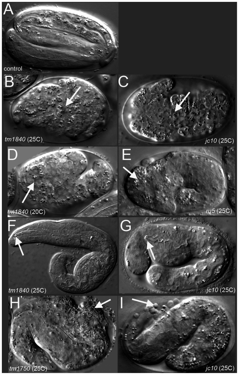
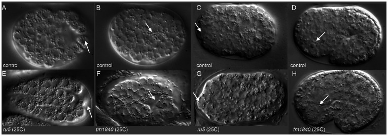
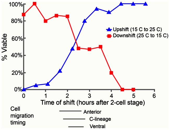
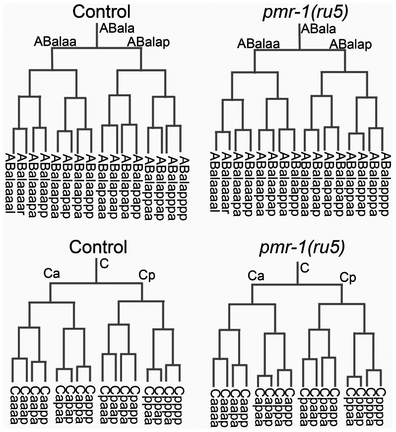
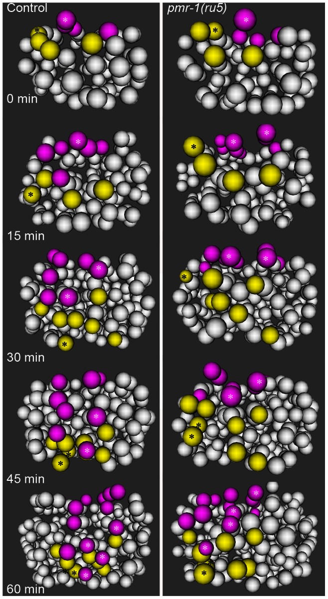
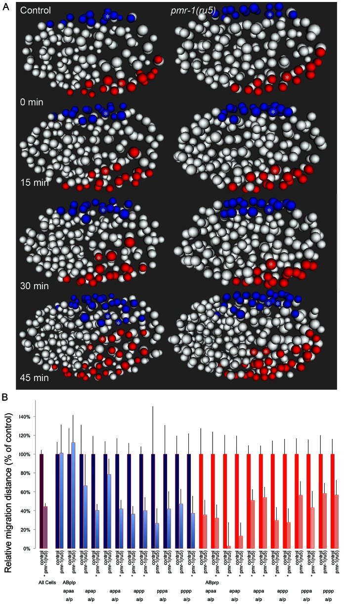
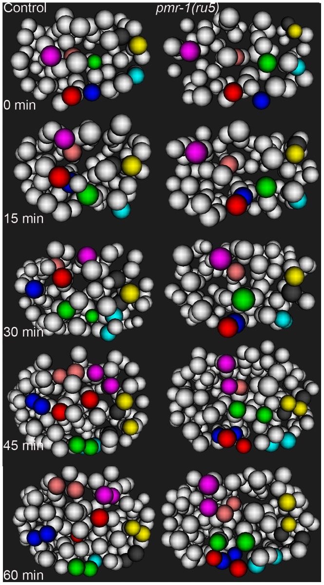
Maintaining levels of calcium in the cytosol is important for many cellular events, including cell migration, where localized regions of high calcium are required to regulate cytoskeletal dynamics, contractility, and adhesion. Studies show inositol-trisphosphate receptors (IP3R) and ryanodine receptors (RyR), which release calcium into the cytosol, are important regulators of cell migration. Similarly, proteins that return calcium to secretory stores are likely to be important for cell migration. The secretory protein calcium ATPase (SPCA) is a Golgi-localized protein that transports calcium from the cytosol into secretory stores. SPCA has established roles in protein processing, metal homeostasis, and inositol-trisphosphate signaling. Defects in the human SPCA1/ATP2C1 gene cause Hailey-Hailey disease (MIM# 169600), a genodermatosis characterized by cutaneous blisters and fissures as well as keratinocyte cell adhesion defects. We have determined that PMR-1, the Caenorhabditis elegans ortholog of SPCA1, plays an essential role in embryogenesis. Pmr-1 strains isolated from genetic screens show terminal phenotypes, such as ventral and anterior enclosure failures, body morphogenesis defects, and an unattached pharynx, which are caused by earlier defects during gastrulation. In Pmr-1 embryos, migration rates are significantly reduced for cells moving along the embryo surface, such as ventral neuroblasts, C-derived, and anterior-most blastomeres. Gene interaction experiments show changing the activity of itr-1/IP3R and unc-68/RyR modulates levels of embryonic lethality in Pmr-1 strains, indicating pmr-1 acts with these calcium channels to regulate cell migration. This analysis reveals novel genes involved in C. elegans cell migration, as well as a new role in cell migration for the highly conserved SPCA gene family.


Conflict of interest statement

The authors have declared that no competing interests exist.

Figures

Figure 1
Figure 1. pmr-1 mutant embryos show a range of terminal phenotypes at the temperatures tested.
(A) Control embryos elongate into the vermiform shape just prior to hatching. In contrast, all four mutant alleles of pmr-1 produce the same range of terminal phenotypes that include B) and C) enclosure defects resulting in full body ruptures (arrow), D) and E) head ruptures (arrow), F) and G) a detached pharynx and body morphogenesis defects (arrow) or H) and I) cells outside the embryo (arrow). Embryos shown include deletion alleles pmr-1(tm1840) (B, D, F) and pmr-1(tm1750) (H), as well as hypomorphic alleles pmr-1(jc10) (C, G, I) and pmr-1(ru5) (E). See Table 2 for specific terminal phenotype frequencies for each allele. In all images, anterior is to the left.
Figure 2
Figure 2. pmr-1 mutant embryos show differences in the positioning of cells during and after gastrulation.
A) In control embryos, the C blastomeres are positioned at the posterior end of the embryo, on the ventral surface, surrounding the gastrulation cleft (arrow). E) In pmr-1 mutant embryos at the same developmental stage, the C blastomeres are shifted to a more dorsal position, out of the ventral focal plane (arrow). B) In control embryos, the gastrulation cleft closes as the last ventral cell ingresses (arrow). F) In pmr-1 mutant embryos at the same stage, the gastrulation cleft remains open (arrow). C) In control embryos, anterior cells form a smooth continuum (arrow), but are displaced in pmr-1 mutant embryos (G, arrow). D) In control embryos, the basement membrane that surrounds the anterior pharynx disappears as these cells migrate to the anterior (arrow, . However, disruption in cell positioning disrupts this process in pmr-1 mutant embryos (H, arrow), preventing pharyngeal attachment. Genotypes are N2 control (A–E), pmr-1(tm1840) (F, H), or pmr-1(ru5) (E, G), all grown at 25°C. In all images, anterior is to the left. A–C and E–G are ventral views; D and H are lateral views.
Figure 3
Figure 3. The temperature-sensitive period for pmr-1(ru5) embryos is during anterior, C-lineage, and ventral cell migrations.
In the upshift experiments, embryos, extracted from gravid adults grown at the permissive temperature (15°C) from the mid-L4 stage, were switched to the restrictive temperature (25°C) at the indicated time. Embryos were then maintained at the restrictive temperature for the duration of embryogenesis and scored “viable” if they thrived past the L1 stage. In the downshift experiments, the same protocol was followed except that the embryos were switched from restrictive temperatures (25°C) to permissive temperatures (15°C) at the indicated times. The lines below the graph correspond to the times when anterior, C-derived, and ventral lineage cell migrations were assayed. All times were normalized to correspond to development at 25°C; n = 5 to 36 embryos at each time point.
Figure 4
Figure 4. Cell division patterns and timing are normal in pmr-1(ru5) embryos.
We determined the cell division patterns in control and pmr-1(ru5) embryos at 25°C to ∼300-cell stage using 4-D microscopy and cell lineage analysis software (Materials and Methods; n = 6 for each strain). Two representative lineages for control (left) and pmr-1(ru5) (right) embryos are shown. We did not observe any differences in cell lineage, cell deaths, or cell division timing in pmr-1(ru5) embryos throughout the temperature-sensitive period. Apoptotic cell deaths, which begin at least one cell division after the temperature sensitive period, occurred normally in all lineages examined (n = 2 controls; n = 4 pmr-1(ru5) strains).
Figure 5
Figure 5. Cell migration defects in the C lineage cells of pmr-1(ru5) embryos.
In control embryos (left panels), the C lineage muscle (yellow) and hypodermal (magenta) precursors migrate from dorsal to ventral positions. The migration rates of control embryos are significantly faster than in pmr-1(ru5) embryos (right panels). The muscle cell precursor Capa and its descendants (yellow; *) and the hypodermal cell precursor Cpap, and its descendants (magenta; *) show the migration and division of a single cell lineage in controls and pmr-1(ru5) mutant embryos. Average rates of migration of these cells for pmr-1(ru5) embryos are 55% of controls for Capa and 40% for Cpap (n = 6 for each strain; p<0.05). Each panel represents a 15-minute interval, starting at 1.8 hours (∼97 cells) after the 2-cell stage at 25°C, which corresponds with the temperature-sensitive period. Posterior view is shown, with dorsal at top.
Figure 6
Figure 6. Cell migration defects in ventral cells of pmr-1(ru5) embryos.
A. Ventral ABp-derived blastomeres migrate from the left (ABplp; blue) or right (ABprp; red) side of the embryo toward the ventral mid-line in control embryos (left panels). In pmr-1(ru5) embryos (right panels), the migration of these blastomeres is significantly reduced. For example, the rectal and interneuron precursor AB.plp appp (Blue; *) and the ring interneuron and ventral cord neuron precursor AB.prp appa (Red; *) migrate further in control than in pmr-1(ru5) embryos. Each panel represents a 15-minute interval, starting at 2 hours (∼162 cells) after the 2-cell stage at 25°C, which corresponds to the temperature-sensitive period. Ventral view is shown, with anterior to the left. B. In comparisons of average migration distance for ABprp (red) and ABplp (blue) blastomeres, cells migrated significantly farther in controls (dark boxes) than in pmr-1(ru5) (light boxes), with significant reductions in migration distance in at least one daughter in 8 of the 12 cells examined. The migration rates in pmr-1(ru5) embryos were 44% of those in controls, when all ventral blastomere migration values were compared (purple). T-test, * indicates p<0.05. Specific lineages tested are indicated. Measurements were taken starting at 2 hours after the 2-cell stage at 25°C (∼162 cells), and again 45 minutes later.
Figure 7
Figure 7. Cell migration defects in the anterior-most cells of pmr-1(ru5) embryos at 25°C.
The anterior cells of control embryos (left panels) undergo distinct and reproducible cell rearrangements during gastrulation. CANL, labial, and ring ganglia precursors ABalapaa (magenta) and ABalaapa (blue) cross the left/right mid-line, labial and ring ganglia precursors ABalpppa (yellow) and ABalppap (gray) cross the dorsal/ventral mid-line and pharyngeal precursor ABalpapp (cyan) migrates from the left to a ventral position. Other cells, such as ring ganglia precursor ABalaaaa (red), migrate from peripheral to central positions or vice versa, as with pharyngeal precursor ABalpaaa (green) and labial/ring ganglia/CANL precursor ABalappa (pink). In a pmr-1(ru5) embryo (right panels), many of these anterior cells migrate shorter distances, migrate in the wrong direction, or fail to change directions, resulting in cells that are mis-positioned. While not every cell showed a migration defect in each embryo, measurements of the Cartesian coordinates for all of these 8 cells except ABalpppa (yellow), as well as three others not labeled above (labial and ring ganglia precursors ABalaaap, ABalpppp, and ABalppaa) showed significant positional differences in pmr-1(ru5) embryos (n = 6) compared to control embryos (n = 6; p<0.05). Each panel represents a 15-minute interval, starting at 1.6 hours (∼88 cells) after 2-cell stage at 25°C, which corresponds to the temperature-sensitive period. Anterior view is shown, with dorsal (top) and embryo right (left). Cell fate information from , .

References

    1. Brini M, Carafoli E (2009) Calcium pumps in health and disease. Phys Rev 89: 1341–1378. - PubMed
    1. Brundage RA, Fogarty KE, Tuft RA, Fay FS (1991) Calcium gradients underlying polarization and chemotaxis of eosinophils. Science 254: 703–706. - PubMed
    1. Ridley AJ, Schwartz MA, Burridge K, Firtel RA, Ginsberg MH, et al. (2003) Cell Migration: Integrating signals from front to back. Science 302: 1704–1709. - PubMed
    1. Wei C, Wang X, Zheng M, Cheng H (2012) Calcium gradients underlying cell migration. Curr Opin Cell Biol 24: 254–261. - PubMed
    1. Wei C, Wang X, Chen M, Ouyang K, Zheng M, et al. (2010) Flickering calcium microdomains signal turning of migrating cells. Can J Physiol Pharmacol 88: 105–110. - PubMed

Publication types

MeSH terms

Substances

LinkOut - more resources