. 2013 May 16;8(5):e63718.
doi: 10.1371/journal.pone.0063718. Print 2013.

Lysophosphatidic acid mediates myeloid differentiation within the human bone marrow microenvironment

Affiliations

Lysophosphatidic acid mediates myeloid differentiation within the human bone marrow microenvironment

Denis Evseenko et al. PLoS One. .

Abstract

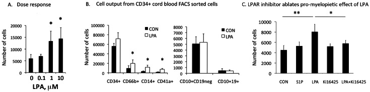
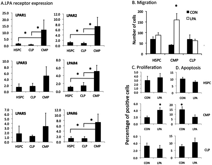
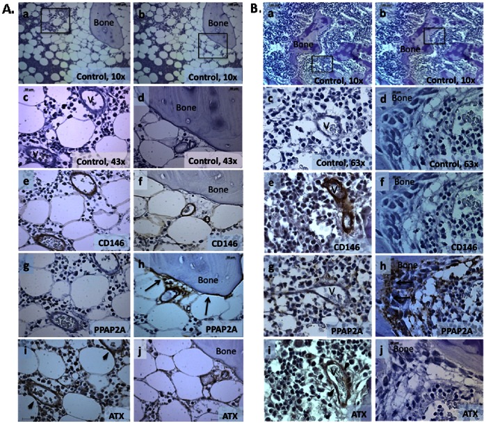
Lysophosphatidic acid (LPA) is a pleiotropic phospholipid present in the blood and certain tissues at high concentrations; its diverse effects are mediated through differential, tissue specific expression of LPA receptors. Our goal was to determine if LPA exerts lineage-specific effects during normal human hematopoiesis. In vitro stimulation of CD34+ human hematopoietic progenitors by LPA induced myeloid differentiation but had no effect on lymphoid differentiation. LPA receptors were expressed at significantly higher levels on Common Myeloid Progenitors (CMP) than either multipotent Hematopoietic Stem/Progenitor Cells (HSPC) or Common Lymphoid Progenitors (CLP) suggesting that LPA acts on committed myeloid progenitors. Functional studies demonstrated that LPA enhanced migration, induced cell proliferation and reduced apoptosis of isolated CMP, but had no effect on either HSPC or CLP. Analysis of adult and fetal human bone marrow sections showed that PPAP2A, (the enzyme which degrades LPA) was highly expressed in the osteoblastic niche but not in the perivascular regions, whereas Autotaxin (the enzyme that synthesizes LPA) was expressed in perivascular regions of the marrow. We propose that a gradient of LPA with the highest levels in peri-sinusoidal regions and lowest near the endosteal zone, regulates the localization, proliferation and differentiation of myeloid progenitors within the bone marrow marrow.


Conflict of interest statement

Competing Interests: The authors have declared that no competing interests exist.

Figures

Figure 1
Figure 1. In vitro system for differentiation of hematopoietic cells.
A. In vitro differentiation of un-fractionated CD34+ progenitors co-cultured on a stromal monolayer in medium containing 5% LPA-depleted serum in the presence of thrombopoietin, Flt3 Ligand and IL-7, cytokines that allow generation of both myeloid (CD45+CD14+ monocytes, CD45+CD66b granulocytes and CD45negCD41a+ megakaryocytes) and B lymphoid cells: CD45+CD10+ CD19neg or CD45+CD10+CD19+. Control panel represents unstained cells. B. Autotaxin protein expression in commonly used stromal lines: MS5, OP9 and human bone marrow-derived mesenchymal stromal cells. Positive signal is shown in brown color (DAB). Magnification 20X. Images were acquired using the Zeiss Axiovision software version 4.8 Carl Zeiss Microscope (Carl Zeiss, Germany).
Figure 2
Figure 2. LPA stimulates generation of myelopoietic lineages from cord blood CD34+ progenitor cells.
A. Dose response of CD34+ cord blood FACS sorted cells to increasing concentration (0.1, 1, 10 uM of LPA). Detection of CD14+ monocytes was used as readout of activity. Mean ± standard deviation (SD), Mean 3 *p<0.05 compared to control cells. B. LPA stimulated generation of myeloid (monocytes, granulocytes and megakaryocytes), but not lymphoid (B-cell) differentiation from CD34+ cells. Freshly sorted CB CD34+ cells were cultured on OP9 stroma for 4 weeks in medium supplemented with 5% LPA-depleted (charcoal treated) serum and growth factor combinations permissive for both myeloid and lymphoid differentiation in the absence (CON = control) or presence of LPA (1 uM). The total number of cells per well in each condition was determined by counting in hematocytometer, and the number of cells of each immunophenotype (shown on the y-axis) was calculated based on % of each lineage phenotype by FACS multiplied to total cell number in each well. Shown is Mean ± standard deviation (SD), N = 4 independent experiments, *p<0.05. C. Stimulatory effects of LPA on myelopoiesis can be ablated using LPA receptor antagonist Ki16425. Myelopoietic differentiation of CD34+ cord blood cells was assessed by the generation of CD14+CD45+ monocytes at 7 days of culture. Concentration of tested compounds: LPA and S1P –1 uM, Ki16425–5 uM. Cord blood samples from 4 donors were analyzed independently and results shown as Mean ± SD. ** P<0.01, * P<0.05. CON = Control.
Figure 3
Figure 3. Myeloid progenitors are functional targets of LPA.
A. LPA receptor mRNA expression in hematopoietic stem-progenitor cells (HSPC), common lymphoid (CLP) and myeloid progenitors (CMP) by qPCR. N = 4 independent experiments; *p<0.05. B. Migration in Transwell® experiments in the presence or absence of 18∶1 Oleoyl-LPA, 12 hours after seeding of HSPC, CMP or CLP. C. Proliferation of each cell type shown measured by 48 hour BrdU uptake. Y axis shows % of BrdU positive cells) D. Apoptosis of each cell type measured by the Annexin V assay (Y axis-shows the % of Annexin V positive cells). Mean ± SD; N = 3 independent experiments; *p<0.05.
Figure 4
Figure 4. Spatial distribution of autotaxin and PPAP2A in adult bone marrow.
A. Adult Bone marrow. Low power (10x) unstained sections showing two different focal plans (a, b). (A–c,e,g,i) shows high power views (43x) of boxed area in (a). (A–d,f,h,j) show high power views of boxed area in (b). (Aa–d) controls stained with secondary antibody only, (A–e, f) CD146 detection of perivascular cells, (A–g, h) PPAP2A expression in endosteal region (arrows), but not perivascular cells, (A–i, j) ATX expression limited to perivascular cells (arrow heads). V = vascular space. B. Fetal Bone marrow. Consistent with the expression pattern of PPAP2A in adult BM, PPAP2A immunoreactivity in fetal BM was predominantly located in bone surfaces lining osteoblasts (arrows), ATX expression limited to perivascular cells (arrow heads) (Fig. 2B–g, h). Low power views (10x) are shown in a, and b. High power views (63x) of areas boxed in panels at left are shown in Bc, e, g, i. High power views (63x) of areas boxed in panels at right are shown in Bd, f, h and j. Positive signals in A and B are shown in brown color (DAB). Images were acquired using the Zeiss Axiovision software version 4.8 Carl Zeiss Microscope (Carl Zeiss, Germany) equipped with ApoTome.2: Modules for Axio Imager.2 and Axio Observer with 40x (1.3 numerical aperture (NA)) and 63x (1.4 NA) oil-immersion objectives.

References

    1. Sano T, Baker D, Virag T, Wada A, Yatomi Y, et al. (2002) Multiple mechanisms linked to platelet activation result in lysophosphatidic acid and sphingosine 1-phosphate generation in blood. J Biol Chem 277: 21197–21206. - PubMed
    1. Tigyi G, Miledi R (1992) Lysophosphatidates bound to serum albumin activate membrane currents in Xenopus oocytes and neurite retraction in PC12 pheochromocytoma cells. J Biol Chem 267: 21360–21367. - PubMed
    1. Tokumura A, Majima E, Kariya Y, Tominaga K, Kogure K, et al. (2002) Identification of human plasma lysophospholipase D, a lysophosphatidic acid-producing enzyme, as autotaxin, a multifunctional phosphodiesterase. J Biol Chem 277: 39436–39442. - PubMed
    1. Blackburn J, Mansell JP (2012) The emerging role of lysophosphatidic acid (LPA) in skeletal biology. Bone 50: 756–762. - PubMed
    1. Choi JW, Herr DR, Noguchi K, Yung YC, Lee CW, et al. (2010) LPA receptors: subtypes and biological actions. Annu Rev Pharmacol Toxicol 50: 157–186. - PubMed

Publication types

MeSH terms