. 2013 May 16;8(5):e63907.
doi: 10.1371/journal.pone.0063907. Print 2013.

HMGB1 accelerates alveolar epithelial repair via an IL-1β- and αvβ6 integrin-dependent activation of TGF-β1

Affiliations

HMGB1 accelerates alveolar epithelial repair via an IL-1β- and αvβ6 integrin-dependent activation of TGF-β1

Jean-François Pittet et al. PLoS One. .

Erratum in

  • PLoS One. 2013;8(10). doi:10.1371/annotation/88f820f2-18dd-4d3b-8989-68f170b26b04

Abstract

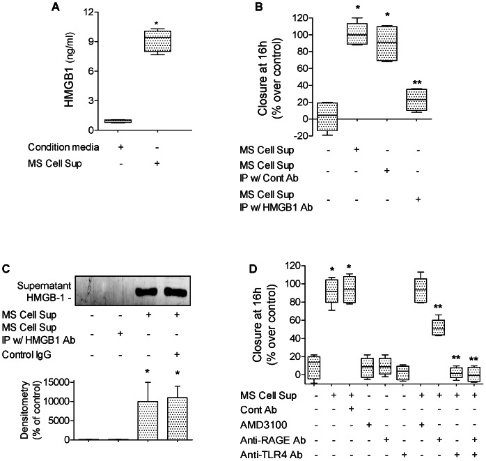
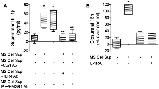
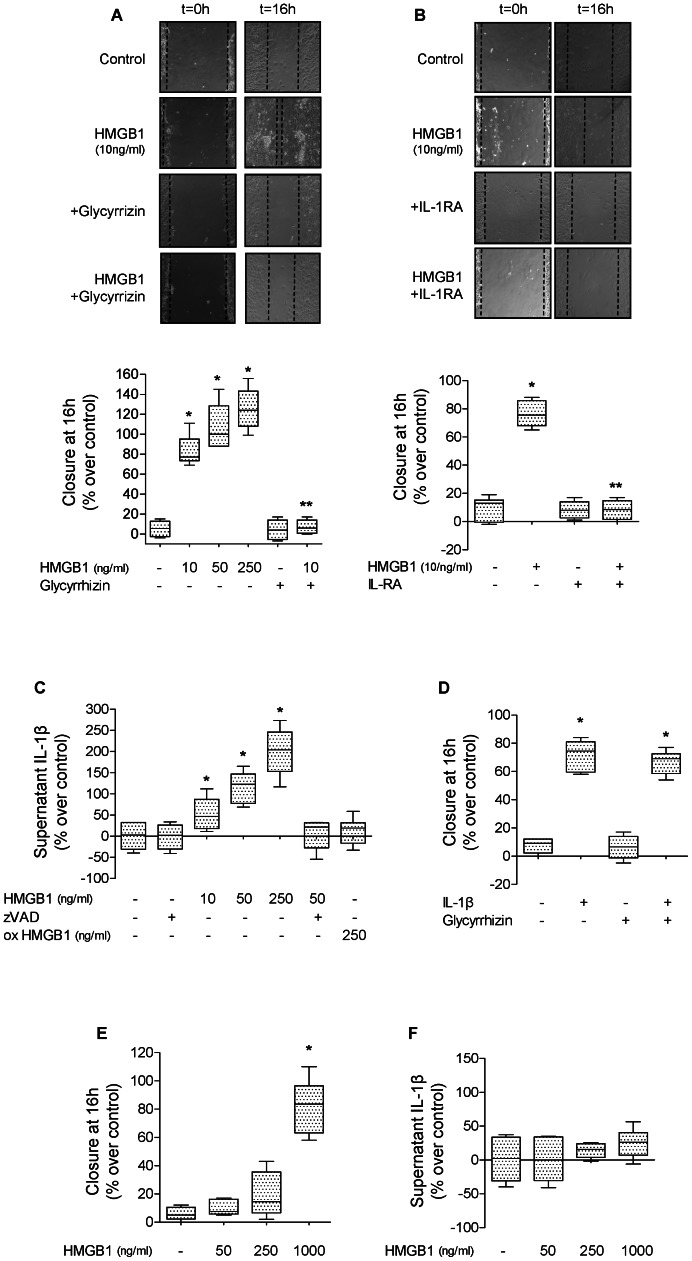
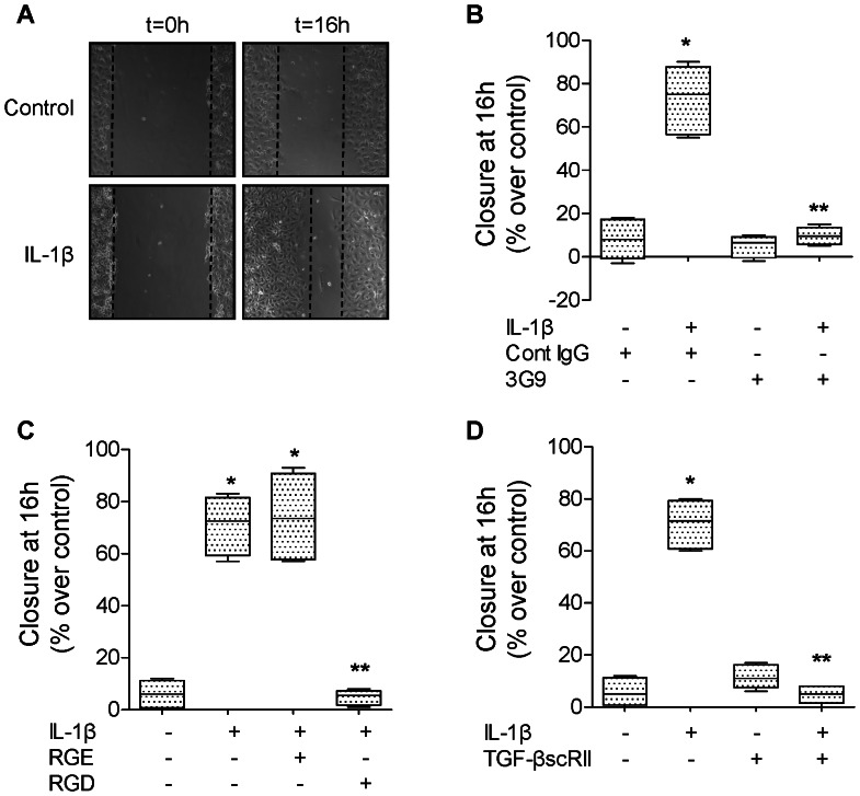
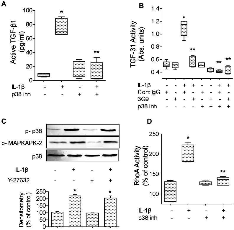
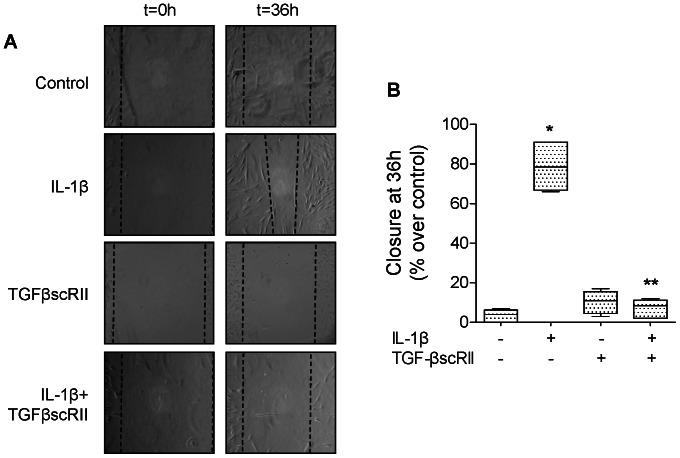
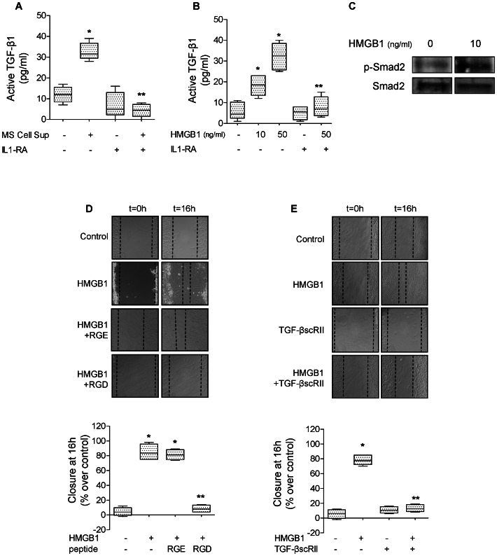
High mobility group box 1 (HMGB1) protein is a danger-signaling molecule, known to activate an inflammatory response via TLR4 and RAGE. HMGB1 can be either actively secreted or passively released from damaged alveolar epithelial cells. Previous studies have shown that IL-1β, a critical mediator acute lung injury in humans that is activated by HMGB1, enhances alveolar epithelial repair, although the mechanisms are not fully understood. Herein, we tested the hypothesis that HMGB1 released by wounded alveolar epithelial cells would increase primary rat and human alveolar type II cell monolayer wound repair via an IL-1β-dependent activation of TGF-β1. HMGB1 induced in primary cultures of rat alveolar epithelial cells results in the release of IL-1β that caused the activation of TGF-β1 via a p38 MAPK-, RhoA- and αvβ6 integrin-dependent mechanism. Furthermore, active TGF-β1 accelerated the wound closure of primary rat epithelial cell monolayers via a PI3 kinase α-dependent mechanism. In conclusion, this study demonstrates that HMGB1 released by wounded epithelial cell monolayers, accelerates wound closure in the distal lung epithelium via the IL-1β-mediated αvβ6-dependent activation of TGF-β1, and thus could play an important role in the resolution of acute lung injury by promoting repair of the injured alveolar epithelium.


Conflict of interest statement

Competing Interests: The authors have declared that no competing interests exist.

Figures

Figure 1
Figure 1. Endogenous HMGB1 released by damaged primary rat ATII cells increases alveolar epithelial wound closure.
(A) HMGB1 was elevated in cell supernatant from rat ATII monolayers that underwent scratch wounds (MS Cell Sup) compared to cell supernatant from rat ATII monolayers that did not undergo scratch wounds (condition media). (B) MS Cell Sup increases the rate of wound closure of primary rat ATII cell monolayers compared to cell supernatant from rat ATII monolayers that did not undergo scratch wounds. HMGB1 was depleted from MS Cell Sup by immunoprecipitation using 30 µg/ml of HMGB1 specific Ab (MS Cell Sup IP w/HMGB1 Ab). Controls were MS Cell Sup immunoprecipitated with a control IgG (MS Cell Sup IP w/Cont Ab). (C) HMGB1 is secreted by primary rat ATII cell monolayers after scratch wounds. Multiple scratches (MS) were performed on primary rat ATII cell monolayers. Fresh cell media were added for 6 hours to the monolayers after extensive washes. Cell supernatants were then centrifuged to remove dead cells and cell debris, then analyzed by western blot (40 µl loaded per lanes from a 1 ml MS Cell Sup sample). (D) MS Cell Sup increases the rate of wound closure of a primary rat ATII cell monolayers via RAGE- and TLR4-dependent pathways, but not via a CXCR4-dependent mechanism. MS Cell Sup, and either 30 µg/ml of blocking RAGE or TLR4 antibodies or their isotype control IgG, or 1 µM of AMD3100, a CXCR4 inhibitor, were added to the monolayers after the scratch. Rate of wound closure is expressed as percent of control 16 h after wounding. *p<0.05 from monolayers exposed to control cell media; **p<0.05 from monolayers exposed to MS Cell Sup. For western blot experiments, one representative experiment is shown, three additional experiments gave comparable results; *p<0.05 from monolayers exposed to condition media.
Figure 2
Figure 2. Endogenous HMGB1 released by primary rat ATII cell monolayers after scratch wounds increases alveolar epithelial wound closure via an IL-1β-dependent mechanism.
(A) Supernatant from primary rat ATII monolayers collected 6 hours after multiple scratches (MS Cell Sup) induces an increase in the secretion of IL-1β by primary rat ATII cell monolayers via a TLR4-dependent pathway. MS Cell Sup, a blocking TLR4 antibody or its isotype control IgG were added to the monolayers after the scratch. HMGB1 was depleted from MS Cell Sup by immunoprecipitation using 30 µg/ml of HMGB1 specific Ab (MS Cell Sup IP w/HMGB1 Ab). Controls were MS Cell Sup immunoprecipitated with a control IgG (MS Cell Sup IP w/Cont Ab). IL-1β was measured by ELISA (see methods) in the cell supernatant. (B) IL-1β receptor antagonist (IL-1RA) prevented the MS Cell Sup-dependent increase in the rate of wound closure of primary rat ATII cell monolayers. MS Cell Sup, and IL-1β receptor antagonist (IL-1RA, 1 µg/ml) or its vehicle were added to the monolayers after the scratch. Rate of wound closure is expressed as percent of control 16 h after wounding. *p<0.05 from monolayers exposed to condition media. **p<0.05 from monolayers exposed to MS Cell Sup.
Figure 3
Figure 3. Human recombinant HMGB1 increases alveolar epithelial wound closure via an IL-1β-dependent signaling pathway.
(A) Human recombinant HMGB1 increases the rate of wound closure of primary rat ATII cell monolayers. HMGB1 (10 ng/ml) or its vehicle was added to the monolayers after scratch. Phase contrast microscopy (20X magnification) immediately after wounding (left panels, t = 0 h) and after 16 h (right panels t = 16 h). Scale bar: 100 µm. In some experiments, glycyrrhizin or its vehicle was added to the monolayers after the scratch. (B) IL-1 receptor antagonist (IL-1RA) prevents HMGB1-dependent increase in rate of wound closure of primary rat ATII cell monolayers. HMGB1 (10 ng/ml) and/or IL-1 receptor antagonist (IL-1RA, 20 µg/ml) or their respective vehicles were added to the monolayers after the scratch. Phase contrast microscopy (20X magnification) immediately after wounding (left panels, t = 0 h) and after 16 h (right panels t = 16 h). Scale bar: 100 µm. (C) HMGB1 increases the secretion of IL-1β by primary rat ATII cell monolayers. HMGB1 (10, 50 and 250 ng/ml, 6 h) or fully oxidized HMGB1 (ox HMGB1, 250 ng/ml) or their respective vehicles was added to the cell monolayers, and IL-1β was measured by ELISA (see methods) in the cell supernatant. In some experiments, zVAD or its vehicle was added to the cell medium 30 min prior to HMGB1. (D) Glycyrrhizin prevents HMGB1-dependent increase in rate of wound closure of primary rat ATII cell monolayers. (E) High dose of Human recombinant HMGB1 (1 µg/ml) is required to increase the rate of wound closure of 3T3 fibroblasts. HMGB1 (50, 250, 1000 ng/ml) or its vehicle was added to the monolayers after scratch. (F) HMGB1 does not increase the secretion of IL-1β by 3T3 fibroblasts. HMGB1 (50, 250 and 1000 ng/ml, 6 h) or its vehicle was added to the cell monolayers, and IL-1β was measured by ELISA (see methods) in the cell supernatant. IL-1β (10 ng/ml) and glycyrrhizin (20 µg/ml) or their respective vehicles were added to the monolayers after the scratch. Rate of wound closure is expressed as percent of control 16 h after wounding. *p<0.05 from monolayers exposed to HMGB1 or IL-1β vehicles. **p<0,005 from monolayers exposed to HMGB1.
Figure 4
Figure 4. IL-1β increases wound closure via αvβ6 integrin and TGF-β1 in primary rat ATII cell monolayers.
(A) IL-1β increases the rate of wound closure of primary rat ATII cell monolayers. IL-1β (10 ng/ml) or its vehicle was added to the monolayers after the scratch. Phase contrast microscopy (20X magnification) immediately after wounding (left panels, t = 0 h) and after 16 h (right panels t = 16 h). Scale bar: 100 µm. (B) A β6 blocking antibody (3G9) prevents IL-1β-dependent increase in rate of wound closure of a primary rat ATII cell monolayer. IL-1β (10 ng/ml). A β6 blocking antibody or its isotype control antibody was added to the monolayers after the scratch. (C) RGD peptides prevent IL-1β-dependent increase in rate of wound closure of a primary rat ATII cell monolayers. IL-1β (10 ng/ml) and RGE or RGD peptide were added to the monolayers after the scratch. (D) A TGF-β1 soluble receptor (TGF-βscRII) prevents IL-1β-dependent increase in rate of wound closure of primary rat ATII cell monolayers. IL-1β (10 ng/ml) and/or TGF-βscRII or their respective vehicles were added to the monolayers after the scratch. Degree of wound closure is expressed as percent of control 16 h after wounding. *p<0.05 from monolayers exposed to IL-1β vehicle. **p<0.05 from monolayers exposed to IL-1β.
Figure 5
Figure 5. IL-1β induces a αvβ6 integrin-dependent activation of TGF-β1 via p38 and RhoA in primary rat ATII cell monolayers.
(A) IL-1β increases active TGF-β1 expression via a p38 MAP kinase-dependent mechanism in primary rat ATII cell monolayers. IL-1β (10 ng/ml, 6 h) or its vehicle was added to the monolayers 30 minutes after pretreatment with a p38 MAP kinase inhibitor (SB202190, 10 µM) or its vehicle. Active TGF-β1 was measured by ELISA, as described in the methods. (B) IL-1β increases TGF-β1 activity via a β6 integrin and p38 MAP kinase-dependent mechanism in primary rat ATII cell monolayers. IL-1β (10 ng/ml, 6 h) or its vehicle was added to the monolayers, 30 minutes after pretreatment with either a β6 blocking antibody, an isotype control antibody, a p38 MAP kinase inhibitor (SB202190, 10 µM) or its vehicle. Active TGF-β1 was measured using mink lung epithelial reporter cells (TMLC, Bioassay for TGF-β1 as described in the methods). (C) RhoA inhibition does not prevent p38 MAP kinase activation by IL-1β in primary rat ATII cell monolayers. IL-1β (10 ng/ml, 10 min) or its vehicle was added to the monolayers 30 minutes after pretreatment with RhoA inhibitor (Y-27632, 10 µM) or its vehicle. (D) A p38 MAP kinase inhibitor (SB202190) prevents RhoA activation by IL-1β in primary rat ATII cell monolayers. IL-1β (10 ng/ml, 30 min) or its vehicle was added to the monolayers, 30 minutes after pretreatment with a p38 MAP kinase inhibitor (SB202190, 10 µM) or its vehicle. *p<0.05 from monolayers exposed to IL-1β vehicle. **p<0.05 from monolayers exposed to IL-1β. For western blot experiments, one representative experiment is shown, three additional experiments gave comparable results; *p<0.05 from monolayers exposed to IL-1β vehicle.
Figure 6
Figure 6. IL-1β increases wound closure via a TGF-β1-dependent mechanism in primary human ATII cell monolayers.
(A) IL-1β increases the rate of wound closure in primary human ATII cell monolayers. IL-1β (10 ng/ml) and/or TGF-βscRII or their respective vehicles were added to the monolayers after the scratch. Phase contrast microscopy (20X magnification) immediately after wounding (left panels, t = 0 h) and after 36 h (right panels t = 36 h). Scale bar: 100 µm. (B) TGF-β1 soluble receptor (TGF-βscRII) prevents IL-1β-dependent increase in rate of wound closure of primary human ATII cell monolayers. IL-1β (10 ng/ml) and/or TGF-βscRII or their respective vehicles were added to the monolayers after the scratch. Rate of wound closure was expressed as percent of control 16 h after wounding. *p<0.05 from monolayers exposed to IL-1β vehicle. **p<0.05 from monolayers exposed to IL-1β.
Figure 7
Figure 7. TGF-β1 increases alveolar epithelial repair via PI3Kα in primary rat ATII cell monolayer.
(A) A β6 blocking antibody (3G9) prevents IL-1β-dependent phosphorylation of Akt in primary rat ATII cell monolayers. IL-1β (10 ng/ml, 30 min) or its vehicle was added to the monolayers 30 minutes after pretreatment with a β6 blocking antibodies or its isotype control antibody (3G9, Cont IgG, 1 µg/ml). (B) TGF-β1 soluble receptor (TGF-βscRII) prevents IL-1β-dependent phosphorylation of Akt in primary rat ATII cell monolayers. IL-1β (10 ng/ml, 30 min) or its vehicle was added to the monolayers 30 minutes after pretreatment with a TGF-β1 soluble receptor (TGF-βscRII, 20 µg/ml) or its vehicle. (C) PI3 kinase inhibition prevents TGF-β1-dependent increase in rate of wound closure of primary rat ATII cell monolayers. TGF-β1 (10 ng/ml) and a broad inhibitor of PI3K (PIK90, 1 µM) or their respective vehicles were added to the monolayers after the scratch. (D) Inhibition of PI3Kα prevents TGF-β1-dependent increase in rate of wound closure of primary rat ATII cell monolayers. TGF-β1 (10 ng/ml) and isoform-specific inhibitors of PI3K (PW12, TGX220, SW14, 0.5 µM) or their respective vehicles were added to the monolayers after the scratch. See Table 1 for PI3K isoform-specific inhibitors IC50. Rate of wound closure is expressed as percent of control 16 h after wounding. *p<0.05 from monolayers exposed to TGF-β1 vehicle. For western blot experiments, one representative experiment is shown, three additional experiments gave comparable results; *p<0.05 from monolayers exposed to IL-1β vehicle. For immunofluorescence experiments, one representative experiment is shown; four additional experiments gave comparable results.
Figure 8
Figure 8. HMGB1 augments alveolar epithelial wound closure via TGF-β and IL-1β in primary rat ATII cells.
(A) Supernatant from primary rat ATII monolayers collected 6 hours after multiple scratches (MS Cell Sup) induces the activation of TGF-β1, which is t blocked by IL-1 receptor antagonist (IL-1RA). MS Cell Sup, and IL-1RA (1 µg/ml) or its vehicle were added to the monolayers for 6 hours. Active TGF-β1 was measured by ELISA, as described in the methods. (B) IL-1 receptor antagonist (IL-1RA) prevents HMGB1-dependent activation of TGF-β1 in primary rat ATII cell monolayers. HGMB1 (10 and 50 ng/ml, 6 h) or its vehicle was added to the monolayers 30 min after pretreatment with IL-1RA or its vehicle. (C) Human recombinant HMGB1 (10 ng/ml, 1 h) causes Smad2/3 phosphorylation in rat primary ATII cell monolayers. (D) RGD peptides prevent HMGB1-dependent increase in rate of wound closure of primary rat ATII cell monolayers. HMGB1 (10 ng/ml) or its vehicle and RGE or RGD peptides (20 µg/ml) were added to the monolayer after the scratch. Phase contrast microscopy (20X magnification) immediately after wounding (left panels, t = 0 h) and after 16 h (right panels t = 16 h). Scale bar: 100 µm. (E) A TGF-β1 soluble receptor (TGF-βscRII) prevents HMGB1-dependent increase in the rate of wound closure of primary rat ATII cell monolayers. HMGB1 (10 ng/ml) and TGF-β1 soluble receptors (TGF-βscRII, 20 µg/ml) or their respective vehicles were added to the monolayers after the scratch. Phase contrast microscopy (20X magnification) immediately after wounding (left panels, t = 0 h) and after 16 h (right panels t = 16 h). Scale bar: 100 µm. Active TGF-β1 was measured by ELISA, as described in the methods. Rate of wound closure is expressed as percent of control 16 h after wounding. *p<0.05 from monolayers exposed to control cell media or HMGB1 vehicle. **p<0.05 from monolayers exposed to MS Cell Sup or HMGB1.

References

    1. Ware LB, Matthay MA (2000) The acute respiratory distress syndrome. N Engl J Med 342: 1334–1349. - PubMed
    1. Geiser T, Jarreau PH, Atabai K, Matthay MA (2000) Interleukin-1beta augments in vitro alveolar epithelial repair. Am J Physiol Lung Cell Mol Physiol 279: L1184–1190. - PubMed
    1. Geiser T, Atabai K, Jarreau PH, Ware LB, Pugin J, et al. (2001) Pulmonary edema fluid from patients with acute lung injury augments in vitro alveolar epithelial repair by an IL-1beta-dependent mechanism. Am J Respir Crit Care Med 163: 1384–1388. - PubMed
    1. Beers MF, Morrisey EE (2011) The three R’s of lung health and disease: repair, remodeling, and regeneration. J Clin Invest 121: 2065–2073. - PMC - PubMed
    1. Chang SW, Feddersen CO, Henson PM, Voelkel NF (1987) Platelet-activating factor mediates hemodynamic changes and lung injury in endotoxin-treated rats. The Journal Of Clinical Investigation 79: 1498–1509. - PMC - PubMed

Publication types

MeSH terms

LinkOut - more resources