. 2013 Jun 11;110(24):9868-72.
doi: 10.1073/pnas.1307864110. Epub 2013 May 22.

General mechanism for modulating immunoglobulin effector function

Affiliations

General mechanism for modulating immunoglobulin effector function

Peter Sondermann et al. Proc Natl Acad Sci U S A. .

Abstract

Immunoglobulins recognize and clear microbial pathogens and toxins through the coupling of variable region specificity to Fc-triggered cellular activation. These proinflammatory activities are regulated, thus avoiding the pathogenic sequelae of uncontrolled inflammation by modulating the composition of the Fc-linked glycan. Upon sialylation, the affinities for Fcγ receptors are reduced, whereas those for alternative cellular receptors, such as dendritic cell-specific intercellular adhesion molecule-3-grabbing nonintegrin (DC-SIGN)/CD23, are increased. We demonstrate that sialylation induces significant structural alterations in the Cγ2 domain and propose a model that explains the observed changes in ligand specificity and biological activity. By analogy to related complexes formed by IgE and its evolutionarily related Fc receptors, we conclude that this mechanism is general for the modulation of antibody-triggered immune responses, characterized by a shift between an "open" activating conformation and a "closed" anti-inflammatory state of antibody Fc fragments. This common mechanism has been targeted by pathogens to avoid host defense and offers targets for therapeutic intervention in allergic and autoimmune disorders.

Keywords: conformational change; sialylated IgG Fc.


Conflict of interest statement

The authors declare no conflict of interest.

Figures
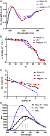
Fig. 1.
Fig. 1.
Sialylation destabilizes the Cγ2 domain of IgG1 Fc. (A) Circular dichroism spectra of IgG1 Fc glycoforms recorded in the far UV range (190–260 nm). Spectra represent an average of four scans with buffer spectra subtracted from sample spectra. (B) Thermal denaturation spectra of Fc glycoforms measured at 206.5 nm. (C) Change in free energy of unfolding, ΔGU, with increasing concentration of chemical denaturant (GnHCl) calculated by the linear extrapolation method. Y-intercept estimates ΔGH2O in aqueous solvent; m value (slope) correlates to solvent-accessible surface area exposed upon unfolding. (D) Fluorescence spectra of sFc and NAse Fc bound to hydrophobic probe ANS.
Fig. 2.
Fig. 2.
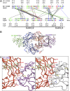
Structural homology of the DC-SIGN/IgG1 Fc and the CD23/IgE Fc complex. (A) Structure-based sequence alignment of human DC-SIGN and IgG1-Fc to the CD23/IgE-Fc complex. The amino acid residues depicted in the one-letter code are colored according to their property (i.e., acidic residues in red, basic in blue, hydrophobic in green). Cysteine residues, all involved in disulfide bridges, are depicted in gray. The interactions between the complex partners are indicated by black lines connecting the involved residues with salt bridges relevant for the CD23/IgE-Fc complex shown in red (7). Hydrogen bonds between IgG and DC-SIGN or CD23 are shown in blue and green lines, respectively. (B) Model of the DC-SIGN/sFc complex. The protein main chains are represented as tubes. The CD23/IgE-Fc complex [thin gray tube, Protein Data Bank (PDB) code 4EZM] is overlaid with the model of the IgG Fc fragment (red and blue) in complex with DC-SIGN (magenta and green). Disulfide bridges and the carbohydrate moiety associated to the Fc fragment are shown as sticks. The square depicts the region shown in detail in C. The generation of the model is described in the supplementary materials. (C) Detailed interaction of DC-SIGN and CD23 with hIgG1-Fc. A close-up of some of the residues expected to be involved in the DC-SIGN/IgG1-Fc (Left) and CD23/IgG1-Fc complex (Right), respectively. The depicted clipping of the complex model is indicated in B.
Fig. 3.
Fig. 3.
Structural changes upon glycosylation of IgG Fc fragments. (A) Schematic view of the principal glycosylation structures attached to Asn297 in the Fc fragment including the respective linkage. Gray shaded parts of the carbohydrate moiety are not displayed in the cartoon. (B) Cartoon of the proposed structural changes within the Fc fragment upon sialylation. The nongalactosylated (G0F) Fc-fragment (top) maintains an open conformation that allows the binding of FcγRs and precludes binding of DC-SIGN or CD23. In the fully α2,6-sialylated Fc (G2FS2), the α1,3-arm (orange/blue) associates with the protein core of the Cγ2 domain, inducing a closed conformation. The resulting closed conformation of the Fc fragment with a changed tertiary structure reveals the binding site for DC-SIGN, whereas that for FcγRs is blocked. The coloring of the carbohydrate moiety is according to the schematic view in A. (C, Upper) Front view of the Fc fragment with the individual domains colored separately with CHOs of the G0F form (PDB code 3AVE). (C, Lower) Front view of the model of fully sialylated Fc (G2FS2) with DC-SIGN bound to it. The coloring of the domains is according to the Fc fragment at top. The right side shows the top view of the respective structures.
Fig. 4.
Fig. 4.
CD23 expressing cells preferentially bind α2,6-sialylated IgG. (A) In cell-based ELISA binding assays, DC-SIGN, CD23, and/or mock transfected CHO-K1 cells were pulsed with an increasing amount of sialylated (sIgG) or asialylated IgG. (A and B) For competition binding assays, DC-SIGN–expressing cells (B) or CD23-expressing cells (C) were pulsed with a constant amount of sIgG and an increasing amount of soluble CD23 or DC-SIGN. In all panels, bound IgG was detected using HRP-labeled anti-human antibody. ECD, extracellular domain.

Comment in

Similar articles

Cited by

References

    1. Sondermann P, Huber R, Oosthuizen V, Jacob U. The 3.2-A crystal structure of the human IgG1 Fc fragment-Fc gammaRIII complex. Nature. 2000;406(6793):267–273. - PubMed
    1. Garman SC, Wurzburg BA, Tarchevskaya SS, Kinet JP, Jardetzky TS. Structure of the Fc fragment of human IgE bound to its high-affinity receptor Fc epsilonRI alpha. Nature. 2000;406(6793):259–266. - PubMed
    1. Anthony RM, Wermeling F, Karlsson MC, Ravetch JV. Identification of a receptor required for the anti-inflammatory activity of IVIG. Proc Natl Acad Sci USA. 2008;105(50):19571–19578. - PMC - PubMed
    1. Hibbert RG, et al. The structure of human CD23 and its interactions with IgE and CD21. J Exp Med. 2005;202(6):751–760. - PMC - PubMed
    1. Anthony RM, Kobayashi T, Wermeling F, Ravetch JV. Intravenous gammaglobulin suppresses inflammation through a novel T(H)2 pathway. Nature. 2011;475(7354):110–113. - PMC - PubMed

Publication types

MeSH terms