

Phenotypic characterization of kidney stone formers by endoscopic and histological quantification of intrarenal calcification
- PMID: 23698231
- PMCID: PMC3784621
- DOI: 10.1038/ki.2013.189
Phenotypic characterization of kidney stone formers by endoscopic and histological quantification of intrarenal calcification
Abstract
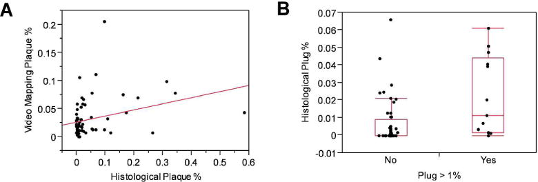
Interstitial Randall's plaques and collecting duct plugs are distinct forms of renal calcification thought to provide sites for stone retention within the kidney. Here we assessed kidney stone precursor lesions in a random cohort of stone formers undergoing percutaneous nephrolithotomy. Each accessible papilla was endoscopically mapped following stone removal. The percent papillary surface area covered by plaque and plug were digitally measured using image analysis software. Stone composition was determined by micro-computed tomography and infrared analysis. A representative papillary tip was biopsied. The 24-h urine collections were used to measure supersaturation and crystal growth inhibition. The vast majority (99%) of stone formers had Randall's plaque on at least 1 papilla, while significant tubular plugging (over 1% of surface area) was present in about one-fifth of patients. Among calcium oxalate stone formers the amount of Randall's plaque correlated with higher urinary citrate levels. Tubular plugging correlated positively with pH and brushite supersaturation but negatively with citrate excretion. Lower urinary crystal growth inhibition predicted the presence of tubular plugging but not plaque. Thus, tubular plugging may be more common than previously recognized among patients with all types of stones, including some with idiopathic calcium oxalate stones.
Figures


Comment in
-
Re: Phenotypic characterization of kidney stone formers by endoscopic and histological quantification of intrarenal calcification.J Urol. 2013 Nov;190(5):1785. doi: 10.1016/j.juro.2013.07.056. Epub 2013 Jul 26. J Urol. 2013. PMID: 24120790 No abstract available.
References
-
- Lieske JC, Pena de la Vega LS, Slezak JM, et al. Renal stone epidemiology in Rochester, Minnesota: An update. Kidney Int. 2006;69:760–764. - PubMed
Publication types
MeSH terms
Substances
Grants and funding
LinkOut - more resources
Full Text Sources
Other Literature Sources
Medical
