. 2013 Aug;87(15):8282-93.
doi: 10.1128/JVI.01102-13. Epub 2013 May 22.

Different requirements for scavenger receptor class B type I in hepatitis C virus cell-free versus cell-to-cell transmission

Affiliations

Different requirements for scavenger receptor class B type I in hepatitis C virus cell-free versus cell-to-cell transmission

Maria Teresa Catanese et al. J Virol. 2013 Aug.

Abstract

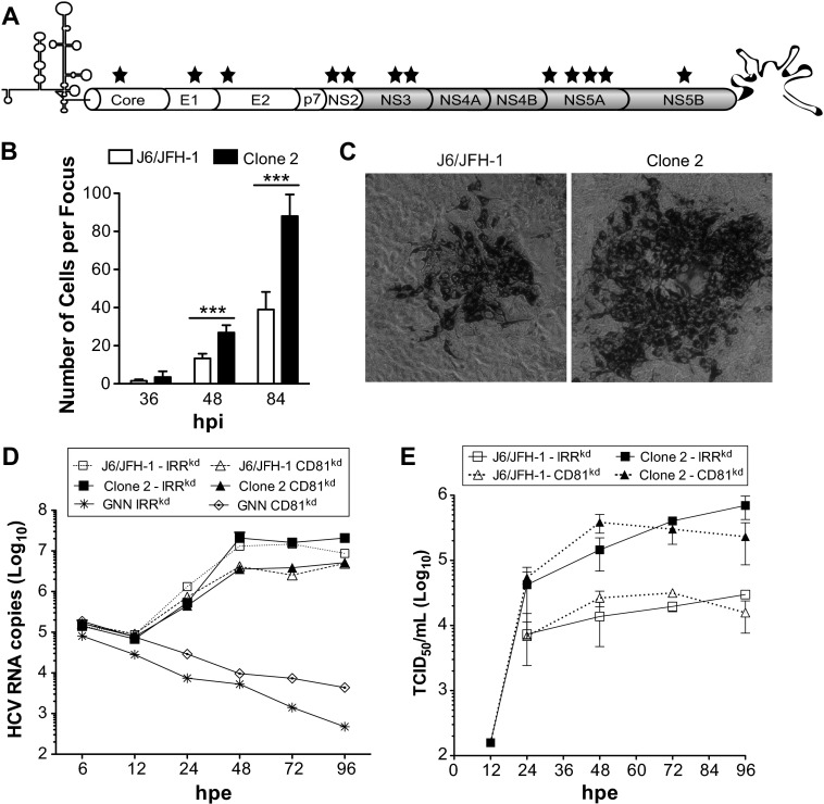
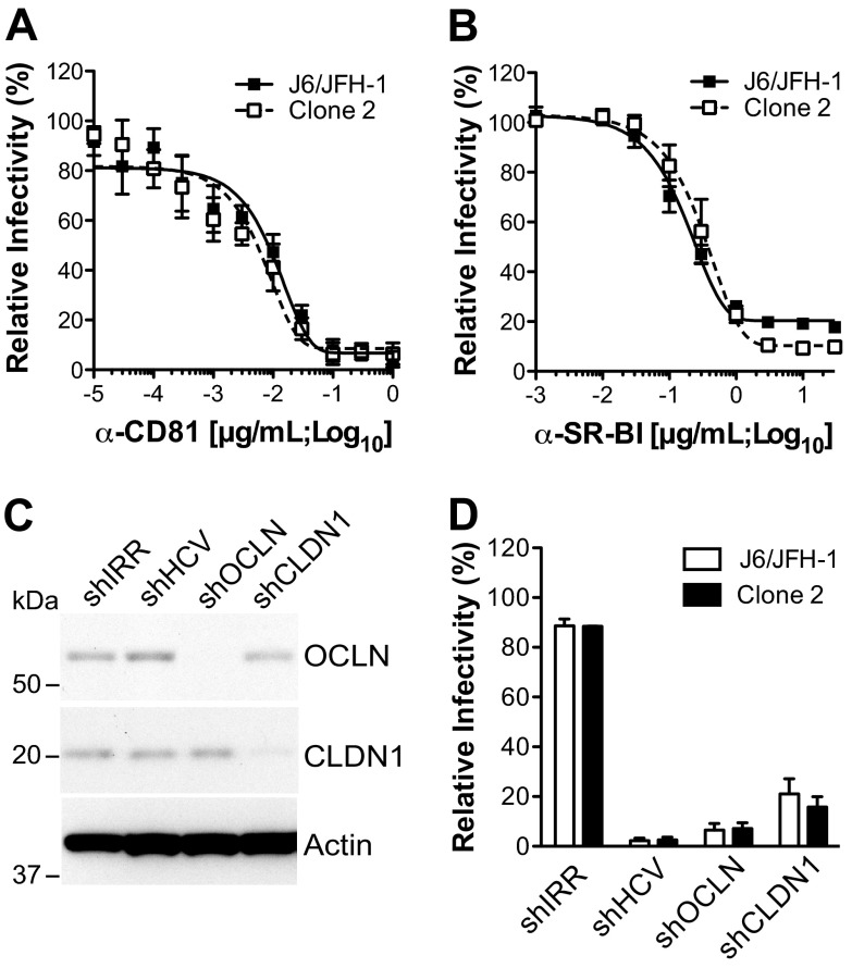
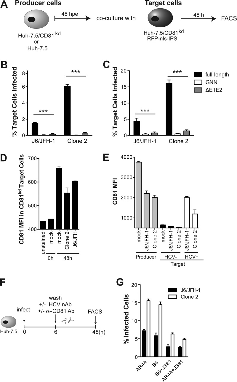
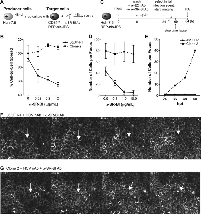
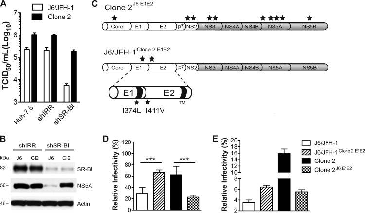
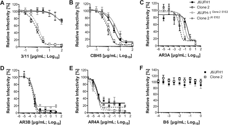
Hepatitis C virus (HCV) is believed to initially infect the liver through the basolateral side of hepatocytes, where it engages attachment factors and the coreceptors CD81 and scavenger receptor class B type I (SR-BI). Active transport toward the apical side brings the virus in close proximity of additional entry factors, the tight junction molecules claudin-1 and occludin. HCV is also thought to propagate via cell-to-cell spread, which allows highly efficient virion delivery to neighboring cells. In this study, we compared an adapted HCV genome, clone 2, characterized by superior cell-to cell spread, to its parental genome, J6/JFH-1, with the goal of elucidating the molecular mechanisms of HCV cell-to-cell transmission. We show that CD81 levels on the donor cells influence the efficiency of cell-to-cell spread and CD81 transfer between neighboring cells correlates with the capacity of target cells to become infected. Spread of J6/JFH-1 was blocked by anti-SR-BI antibody or in cells knocked down for SR-BI, suggesting a direct role for this receptor in HCV cell-to-cell transmission. In contrast, clone 2 displayed a significantly reduced dependence on SR-BI for lateral spread. Mutations in E1 and E2 responsible for the enhanced cell-to-cell spread phenotype of clone 2 rendered cell-free virus more susceptible to antibody-mediated neutralization. Our results indicate that although HCV can lose SR-BI dependence for cell-to-cell spread, vulnerability to neutralizing antibodies may limit this evolutionary option in vivo. Combination therapies targeting both the HCV glycoproteins and SR-BI may therefore hold promise for effective control of HCV dissemination.


Figures

Fig 1
Fig 1
Increased cell-to-cell spread of cell culture-adapted HCV. (A) Schematic representation of the J6/JFH-1-derived clone 2 genome with the locations of mutations indicated (stars). (B) Sizes of J6/JFH-1 and clone 2 foci formed at 36, 48, and 84 hpi on Huh-7.5 cells in the presence of HCV nAb (AR4A; 0.1 μg/ml). Means and standard deviations (SDs) from three independent experiments are shown, and values that were significantly different are indicated (***, P < 0.001). (C) Representative images of J6/JFH-1 and clone 2 foci. Huh-7.5 cells were infected and stained with anti-NS5A antibody (9E10) at 84 hpi. (D, E) Kinetics of RNA replication (D) and production of infectious virus (E) following electroporation of J6/JFH-1 and clone 2 genomes into Huh-7.5 cells encoding an irrelevant shRNA (IRRkd) or CD81kd.
Fig 2
Fig 2
Receptor requirements for cell-free infectivity with J6/JFH-1 and clone 2. (A, B) J6/JFH-1 or clone 2 cell-free infection efficiency in the presence of anti-CD81 (A) or anti-SR-BI (B) mAb. Huh-7.5 cells were preincubated with the indicated concentrations of mAb (μg/ml; log10) and infected with HCVcc in the presence of the mAb. The percentage of HCV-infected cells at 48 hpi was measured by flow cytometry using Alexa Fluor 647-conjugated anti-NS5A antibody (9E10-AF647). MFIs obtained with isotype-matched controls were subtracted. Curves were extrapolated by fitting experimental data with GraphPad Prism (version 5.0) software. (C) Immunoblot for OCLN, CLDN1, and β-actin expression in Huh-7.5 cells expressing shIRR, shHCV, shOCLN, and shCLDN1. Cells were harvested at 72 h posttransduction, and 30 μg total cell lysate was loaded per lane. (D) Huh-7.5 cells expressing the indicated shRNAs were inoculated with J6/JFH-1 or clone 2 at an MOI of 0.1, and HCV infection frequency was measured at 48 hpi at the flow cytometer by staining the cells for NS5A with 9E10-AF647. The value for parental Huh-7.5 cells was set to 100%. Mean values and SDs from three independent experiments are plotted.
Fig 3
Fig 3
Effect of CD81 levels on HCV cell-to-cell transmission. (A) Schematic of coculture experiments. Producer cells (Huh-7.5/CD81kd or Huh-7.5) were cultured for 48 h after electroporation with HCV genomes before mixing with naive Huh-7.5/CD81kd/RFP-nls-IPS target cells at a 1.5:1 ratio. Cocultures were analyzed by flow cytometry 48 h later, after staining with anti-NS5A Ab. The NS5A+ cells in the RFP+ population represented infected target cells. (B, C) NS5A expression in the target cell population was analyzed by FACS to quantify HCV cell-to-cell spread. Graphs show the percentage of infected target cells when cocultured with either Huh7.5/CD81kd (B) or Huh-7.5 (C) producer cells. Full-length, polymerase-defective (GNN), and virus assembly-impaired genomes (ΔE1E2) were tested in parallel. Means and SDs from two independent experiments are shown, and values that differ significantly from the values for HCV spread obtained in the presence of the full-length genomes are indicated. ***, P < 0.001. (D) Cell surface CD81 expression on Huh-7.5/CD81kd target cells was measured by FACS prior to and at 48 h after cultivation with uninfected (mock) or J6/JFH-1- or clone 2-infected Huh-7.5 cells. MFIs of anti-CD81–APC antibody and SDs from two independent experiments are shown. (E) Cocultures between uninfected (mock) or J6/JFH-1- or clone 2-infected Huh-7.5 and naive Huh-7.5/CD81kd/RFP-nls-IPS target cells were permeabilized 48 h later and costained with anti-NS5A/AF488 and anti-CD81–APC antibodies. MFIs obtained with anti-CD81–APC in the CD81+ producer (RFP) and CD81kd target (RFP+) populations are plotted. Target cells were further classified HCV negative (HCV; black bars) and HCV+ (white bars) on the basis of the staining with anti-NS5A/AF488. (F) Schematic of HCV cell-to-cell spread experiments in CD81+ cultures in the presence or absence of HCV-neutralizing and anti-CD81 antibodies. (G) Percentage of infected cells obtained with the two viruses when cultures were treated postinfection (MOI, 0.1) with the indicated single antibody or combinations (each at 1 μg/ml).
Fig 4
Fig 4
Impact of anti-SR-BI antibody on J6/JFH-1 and clone 2 cell-to-cell spread kinetics. (A) Schematic of coculture of HCV-infected Huh-7.5 producer cells with Huh7.5/CD81kd target reporter cells. Producer cells were preinfected with J6/JFH-1 or clone 2 virus for 48 h before mixing at a 1.5:1 ratio with uninfected Huh7.5/CD81kd/RFP-nls-IPS target cells and adding anti-SR-BI antibody. Cocultures were incubated for an additional 48 h and then analyzed by flow cytometry. (B) The percentage of target infected cells at increasing concentrations of SR-BI mAb (C167; 0.02, 0.2, and 2 μg/ml) was measured by FACS by gating on NS5A+/RFP+ double-positive cells. Data are expressed as the percentage of cell-to-cell spread relative to that of an isotype-matched irrelevant control antibody. Mean values and SDs from three independent experiments are plotted. (C) Schematic of HCV focus-forming assay. Huh-7.5 cells stably expressing RFP-nls-IPS were infected with J6/JFH-1 or clone 2 at a low MOI (0.01) for 6 h prior to removal of the inoculum and addition of imaging medium containing anti-E1-E2 (AR4A; 0.1 μg/ml) and anti-SR-BI (C167; 0.1, 1, and 10 μg/ml) antibodies. Time-lapse live-cell imaging of viral spread was started at 24 hpi, when single infected cells could be visualized by nuclear translocation of RFP, and stopped at 69 hpi. Parallel cultures were fixed and analyzed by fluorescence microscopy at 84 hpi to quantify the endpoint size of HCV foci. (D) The size of HCV foci was determined by scoring the number of RFP-positive nuclei in PFA-fixed cell monolayers at 84 hpi. The mean size and SD of 30 distinct HCV-infected cell clusters from three independent experiments are shown. (E) The number of HCV-positive cells over time from representative time-lapse live-cell imaging (see also panels F and G and Videos S1 and S2 in the supplemental material) is shown. RFP-positive nuclei from the same imaging stage position were counted at the indicated time points (hpi). (F, G) Montages of images taken from selected time points beginning at 24 hpi show the formation of J6/JFH-1 and clone 2 foci, respectively, with the highest anti-SR-BI antibody concentration tested (10 μg/ml). Images were captured every 30 min to up to 69 hpi. RFP fluorescence is shown in gray scale. Times (h) from the start of infection are shown on the top left corner, and the white arrows indicate the initially selected HCV-infected cells. See Videos S1 and S2 in the supplemental material for the full time course.
Fig 5
Fig 5
E1 and E2 mutations in clone 2 influence SR-BI use in cell-to-cell spread. (A) The dependence of J6/JFH-1 and clone 2 on SR-BI for propagation in cell culture was assessed by titrating the same viral supernatants on Huh-7.5 wild-type cells or Huh-7.5 cells expressing shIRR or shSR-BI. At 3 days postinoculation, virus titers (TCID50/ml) were determined. Means and SDs from two independent assays are plotted. (B) Immunoblot for SR-BI, NS5A, and β-actin in Huh-7.5 cells expressing shIRR or shSR-BI infected with either J6/JFH-1 (J6) or clone 2 (Cl2) viruses at 72 hpi. Whole-cell lysates (30 μg per lane) were loaded on a 4 to 12% bis-Tris NuPage gel. (C) Schematic representation of the chimeric viruses clone 2J6 E1E2, harboring parental J6/JFH-1 E1-E2 sequences in a clone 2 background, and J6/JFH-1clone 2 E1E2, a J6/JFH-1 genome bearing the clone 2 I374L (E1) and I411V (E2) mutations. Stars, approximate positions of the acquired mutations on the polyprotein. (D) Huh-7.5 cells were transduced with lentiviruses encoding shIRR or shSR-BI and challenged 48 h later with HCV (clone 2, J6/JFH-1, or chimeric viruses). At 3 days postinfection, cells were stained for NS5A and analyzed by FACS. Data are expressed as the percentage of shSR-BI-infected cells relative to the number of cells infected with the shIRR counterpart (set as 100%). Mean values and SDs from three independent experiments are shown, and values that differ significantly are indicated. ***, P < 0.001. (E) Huh-7.5 cells were electroporated with the indicated genomes and used as producers in cocultures with Huh-7.5/CD81kd/RFP-nls-IPS/SR-BIkd/GFP target cells. The graph shows the percentage of infected cells in the double CD81kd/SR-BIkd target population. Means and SDs from two independent experiments are plotted.
Fig 6
Fig 6
Mutations in clone 2 E1 and E2 affect sensitivity to cell-free antibody-mediated neutralization. J6/JFH-1, clone 2, and the chimeric viruses clone 2J6 E1E2 and J6/JFH-1clone 2 E1E2 were preincubated with increasing concentrations (μg/ml) of HCV-specific antibodies 3/11 (A), CBH-5 (B), AR3A (C), AR3B (D), or AR4A (E) or with a human anti-HIV antibody, B6, that served as a negative control (F). The residual virus infectivity at the indicated antibody concentrations (the logarithm of the concentration of antibody is plotted on the x axis) was calculated at 3 days postinfection by NS5A staining and flow cytometric analysis. Infectivity without antibody was set to 100%. Mean values and SDs from four independent experiments are plotted.

References

    1. Lavanchy D. 2011. Evolving epidemiology of hepatitis C virus. Clin. Microbiol. Infect. 17:107–115 - PubMed
    1. Zeisel MB, Cosset FL, Baumert TF. 2008. Host neutralizing responses and pathogenesis of hepatitis C virus infection. Hepatology 48:299–307 - PubMed
    1. Andre P, Komurian-Pradel F, Deforges S, Perret M, Berland JL, Sodoyer M, Pol S, Brechot C, Paranhos-Baccala G, Lotteau V. 2002. Characterization of low- and very-low-density hepatitis C virus RNA-containing particles. J. Virol. 76:6919–6928 - PMC - PubMed
    1. Nielsen SU, Bassendine MF, Burt AD, Martin C, Pumeechockchai W, Toms GL. 2006. Association between hepatitis C virus and very-low-density lipoprotein (VLDL)/LDL analyzed in iodixanol density gradients. J. Virol. 80:2418–2428 - PMC - PubMed
    1. Maillard P, Huby T, Andreo U, Moreau M, Chapman J, Budkowska A. 2006. The interaction of natural hepatitis C virus with human scavenger receptor SR-BI/ClaI is mediated by ApoB-containing lipoproteins. FASEB J. 20:735–737 - PubMed

Publication types

MeSH terms

LinkOut - more resources