. 2013 Aug;87(15):8272-81.
doi: 10.1128/JVI.00969-13. Epub 2013 May 22.

In vivo bioluminescent imaging of influenza a virus infection and characterization of novel cross-protective monoclonal antibodies

Affiliations

In vivo bioluminescent imaging of influenza a virus infection and characterization of novel cross-protective monoclonal antibodies

Nicholas S Heaton et al. J Virol. 2013 Aug.

Abstract

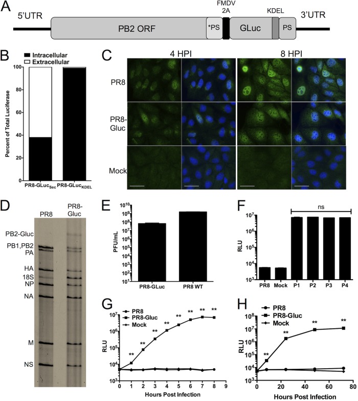
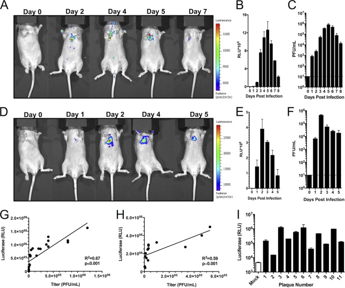
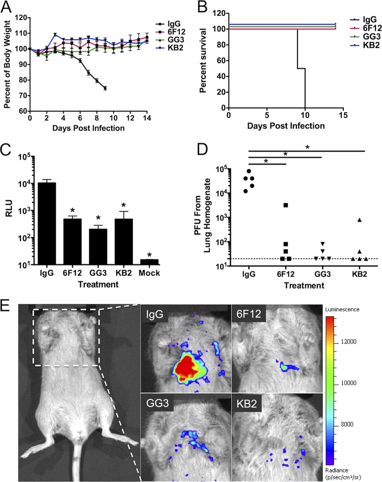
Influenza A virus is a major human pathogen responsible for seasonal epidemics as well as pandemic outbreaks. Due to the continuing burden on human health, the need for new tools to study influenza virus pathogenesis as well as to evaluate new therapeutics is paramount. We report the development of a stable, replication-competent luciferase reporter influenza A virus that can be used for in vivo imaging of viral replication. This imaging is noninvasive and allows for the longitudinal monitoring of infection in living animals. We used this tool to characterize novel monoclonal antibodies that bind the conserved stalk domain of the viral hemagglutinin of H1 and H5 subtypes and protect mice from lethal disease. The use of luciferase reporter influenza viruses allows for new mechanistic studies to expand our knowledge of virus-induced disease and provides a new quantitative method to evaluate future antiviral therapies.


Figures

Fig 1
Fig 1
In vitro characterization of PR8-GLuc. (A) Schematic representation of the PB2 segment encoding the GLuc transgene. *PS represents silent mutations of the original packaging signal; PS represents the duplicated original packaging sequence. (B) MDCK cells were infected with PR8-GLuc with and without a KDEL ER retention signal. The relative percentages of secreted and intracellular GLuc signals are shown. (C) MDCK cells were infected with WT PR8 or PR8-GLuc at an MOI of 1 and fixed at the indicated times. PB2 cellular localization was probed with an anti-PB2 antibody. Scale bar, 20 μm. (D) Viral RNA was extracted from either WT PR8 or PR8-GLuc virions and resolved on a polyacrylamide gel. RNA was stained via silver stain. 18S represents rRNA contamination of the virion preparation. (E) Titers in PFU/ml of WT PR8 and PR8-GLuc after 48 h of growth in eggs at 37°C. (F) MDCK cells were mock infected or infected with WT PR8 or with PR8-GLuc from serial passage experiments in eggs (passages 1 to 4) at an MOI of 1. At 6 hpi cells were harvested, and luciferase assays were performed. (G) MDCK cells were mock or virus infected at an MOI of 1 and incubated without TPCK-trypsin for the indicated time points. At the indicated times, luciferase activity was determined. (H) MDCK cells were mock or virus infected at an MOI of 0.001 and incubated with TPCK-trypsin for the indicated times. Cellular lysates were collected, and luciferase activity was determined. **, P ≤ 0.001; ns, not significant. NA, neuraminidase; RLU, relative light units.
Fig 2
Fig 2
PR8-GLuc infection induces lethal disease in mice and exhibits the expected tropisms. (A and B) DBA/2 mice were infected with the indicated inoculum of PR8-GLuc and monitored for weight loss and lethality. (C and D) BALB/c mice were infected with the indicated inoculum of PR8-GLuc and monitored for weight loss and lethality. (E) DBA/2 mice were infected with 103 PFU of PR8-GLuc or mock infected. Three days after infection, the indicated organs were collected, and the levels of luciferase in these organs were determined (**, P ≤ 0.001). OB, olfactory bulb.
Fig 3
Fig 3
PR8-GLuc infection allows for in vivo imaging. Mice were infected with 103 PFU (A) or 105 PFU (D) and imaged at the indicated days. At the indicated times, lungs were collected from infected animals, and the amounts of luciferase and viral titers were determined. (B and C) Data correspond to the time course for the dose of 103 PFU. (E and F) Data correspond to the time course for the dose of 105 PFU. (G and H) Luciferase levels and viral titers from individual animals were plotted against each other from time courses of doses of 103 PFU(G) and 105 PFU (H). The R2 and P values for the linear regression analyses are indicated on each graph. (I) Lung homogenates from animals at the day 4 time point (inoculum, 105 PFU) were plaque purified. Virus was isolated from each plaque and used to infect MDCK cells for 16 h. Cellular lysates were collected and subjected to luciferase assays. The white bar indicates mock-infected controls, and each black bar indicates the average luciferase value for an individual plaque-purified viral clone.
Fig 4
Fig 4
Novel monoclonal antibodies against the HA stalk region restrict a panel of H1 and H5 viruses. (A) Monoclonal antibodies GG3 and KB2 bind to HA on the surface of infected cells from all tested influenza A H1 virus strains, similar to the previously published pan-H1 antibody 6F12. GG3 and KB2, however, also bind an H5 avian isolate. None of the tested antibodies bound to H3 isolates. (B to E) Monoclonal antibodies GG3 and KB2 were incubated with hemagglutinin from the indicated viruses (as noted on the y axes) expressed and purified via a baculovirus expression system. Plates were coated with the purified protein, and reactivity of GG3 or KB2 via ELISA was determined. Positive (+) sera, polyclonal sera from convalescent mice infected with the appropriate viruses. (F and G) GG3 and KB2 were incubated with the indicated viruses in a plaque reduction assay. (H and I) ELISA plates were coated with hemagglutinin from the H1 PR8 virus or a chimeric HA protein consisting of an H6 head with the group 1 stalk from PR8 (cH6/1). The following antibodies were tested for reactivity against those proteins: PY102 (an H1 head-specific antibody), H6mAb (an antibody specific for H6 head domains), KB2, GG3, and a negative control. Viruses are abbreviated as follows: NY09, influenza A/NewYork/18/2009; Sol06, influenza/A/Solomon Islands/03/2006; VN04, influenza A/Vietnam/1203/2004 (H5N1); Cal09, influenza A/California/04/2009; USSR77, influenza A/USSR/77; TX91, influenza A/Texas/36/91; Brisbane07, influenza A/Brisbane/59/2007; HK68, influenza A/Hong Kong/1/1968; FM47, influenza A/Fort Monmouth/1/1947; N. Cal99 or NC99, influenza A/NewCaledonia/20/1999. Ab, antibody; OD490, optical density at 490 nm.
Fig 5
Fig 5
GG3 and KB2 restrict PR8-GLuc replication in vivo. (A and B) DBA/2 mice were treated i.p. with 5 mg/kg of the indicated antibodies or an isotype control at 2 h preinfection. Mice were then challenged with 5 LD50s of PR8-GLuc and monitored over time for weight loss and survival. (C and D) Lungs from mice infected as described for panels A and B were collected 5 days postinfection and assayed for luciferase levels (C) and viral titers (D). The dotted line in panel D indicates the limit of detection. *, P ≤ 0.05. (E) Mice infected as described for panels A and B were imaged at 5 days postinfection to determine virus replication.
Fig 6
Fig 6
GG3 and KB2 protect BALB/c mice against lethal challenge with pandemic H1 and avian H5 influenza viruses. Mice were administered the indicated doses of antibody GG3 or KB2 and challenged with the pandemic H1N1 isolate NL09 (influenza A/Netherlands/602/2009) or the avian H5N1 isolate rVN04 (influenza A/Vietnam/1203/2004), as indicated. Weight loss and survival were monitored.

Similar articles

Cited by

References

    1. Palese P, Shaw ML. 2007. Orthomyxoviridae: the viruses and their replication, p 1647–1689 In Knipe DM, Howley PM. (ed), Fields virology, 5th ed Lippincott Williams & Wilkins, Philadelphia, PA
    1. Neumann G, Noda T, Kawaoka Y. 2009. Emergence and pandemic potential of swine-origin H1N1 influenza virus. Nature 459:931–939 - PMC - PubMed
    1. Taubenberger JK, Morens DM. 2008. The pathology of influenza virus infections. Annu. Rev. Pathol. 3:499–522 - PMC - PubMed
    1. Kuiken T, Riteau B, Fouchier RA, Rimmelzwaan GF. 2012. Pathogenesis of influenza virus infections: the good, the bad and the ugly. Curr. Opin. Virol. 2:276–286 - PubMed
    1. Close DM, Xu T, Sayler GS, Ripp S. 2011. In vivo bioluminescent imaging (BLI): noninvasive visualization and interrogation of biological processes in living animals. Sensors (Basel) 11:180–206 - PMC - PubMed

Publication types

MeSH terms