. 2013 Aug;87(15):8327-41.
doi: 10.1128/JVI.01025-13. Epub 2013 May 22.

Role of the phosphatidylserine receptor TIM-1 in enveloped-virus entry

Affiliations

Role of the phosphatidylserine receptor TIM-1 in enveloped-virus entry

Sven Moller-Tank et al. J Virol. 2013 Aug.

Abstract

The cell surface receptor T cell immunoglobulin mucin domain 1 (TIM-1) dramatically enhances filovirus infection of epithelial cells. Here, we showed that key phosphatidylserine (PtdSer) binding residues of the TIM-1 IgV domain are critical for Ebola virus (EBOV) entry through direct interaction with PtdSer on the viral envelope. PtdSer liposomes but not phosphatidylcholine liposomes competed with TIM-1 for EBOV pseudovirion binding and transduction. Further, annexin V (AnxV) substituted for the TIM-1 IgV domain, supporting a PtdSer-dependent mechanism. Our findings suggest that TIM-1-dependent uptake of EBOV occurs by apoptotic mimicry. Additionally, TIM-1 enhanced infection of a wide range of enveloped viruses, including alphaviruses and a baculovirus. As further evidence of the critical role of enveloped-virion-associated PtdSer in TIM-1-mediated uptake, TIM-1 enhanced internalization of pseudovirions and virus-like proteins (VLPs) lacking a glycoprotein, providing evidence that TIM-1 and PtdSer-binding receptors can mediate virus uptake independent of a glycoprotein. These results provide evidence for a broad role of TIM-1 as a PtdSer-binding receptor that mediates enveloped-virus uptake. Utilization of PtdSer-binding receptors may explain the wide tropism of many of these viruses and provide new avenues for controlling their virulence.


Figures

Fig 1
Fig 1
TIM-1 threaded on mTIM-1 crystal structure. (A) Amino acid sequences of TIM-1, mTIM-1, TIM-3, and TIM-4 IgV domains aligned using the Clustal W software program. Residues in the TIM-1 PtdSer binding pocket that were mutated are shown in bold. β-Sheets are marked above the alignment with lines and corresponding letters. Residue numbering is based on the TIM-1 sequence. (B) Structures of mTIM-1 IgV (20R8) (left) and a threaded model of TIM-1 IgV (right). β-Sheets are labeled with their corresponding letters, assigned by Santiago et al. (40). (C) PtdSer binding residues of mTIM-1 (above) and TIM-1 (below) are shown on each structure. (D) All residues in the TIM-1 IgV domain mutated in this study are highlighted in black.
Fig 2
Fig 2
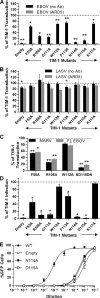
Identification of TIM-1 IgV residues that impact EBOV GP-dependent entry. (A to C) Relative virus transduction into HEK 293T cells mediated by mutant TIM-1 constructs compared to that with WT TIM-1. At 48 h, transfected cells were transduced with EBOV GP-VSVΔG (A), LASV GPC-VSVΔG (B), MARV GP-VSVΔG (C), or full-length EBOV-VSVΔG (C) pseudovirions. (A and B) Transductions were also done in the presence or absence of anti-human TIM-1 MAb ARD5 (1.7 μg/ml). EGFP expression was assessed 24 h later. (D) EBOV GP/rVSV-EGFP infection mediated by TIM-1 constructs in HEK 293T cells (MOI of 1 as determined from titers in Vero cells) relative to WT TIM-1. (E) Serial dilution of EBOV GP/rVSV-EGFP on TIM-1-transfected HEK293T cells. EGFP expression was assessed 24 (A to C) or 48 (D and E) h later by flow cytometry. Data are shown as means ± SEM for at least three replicates. (A to D) Significance was calculated using one-sample t-test comparison to 100 (**, P < 0.001; *, P < 0.01). Significance for all ARD5 transductions for EBOV was defined as P < 0.001.
Fig 3
Fig 3
TIM-1-mediated transduction requires PtdSer binding. (A) Transduction of TIM-1+ H3 cells with EBOV pseudovirions in the presence of increasing concentrations of PtdSer or PtdChl liposomes. (B) Transduction of EBOV and LASV pseudovirions into Vero cells in the presence of increasing EGTA concentrations. EGTA in PBS plus 10% FBS was incubated on Vero cells for 1 h before pseudovirions were added for 4 h. Medium was replaced, and transduction was assessed after 24 h. (C) Transduction of EBOV pseudovirions into empty-vector- or TIM-4-transfected HEK 293T cells relative to TIM-1 transduction. (D) AnxV binds to EBOV pseudovirions. Increasing concentrations of HA-tagged AnxV were incubated with ELISA plates prebound with EBOV pseudovirions (filled) or untreated (open). (E) HEK 293T supernatants containing HA-tagged soluble Axl, mSAP, TIM-1, TIM-4, or mutants of TIM-1, N114D or D115N, were incubated with ELISA plates prebound with EBOV pseudovirions or not treated. Relative protein amounts present in supernatants are shown below in a representative Western blot using anti-HA antisera. (F) Binding of TIM-1 and N114D from supernatants to ELISA plates prebound with PtdSer (filled) or PtdChl (open) liposomes (50 μM). (G) Binding of HA-tagged TIM-1 in the presence of IgG2a, MAb ARD5, MAb A6G2, PtdSer, or PtdChl liposomes or 2 mM EGTA to ELISA plates prebound with EBOV pseudovirions. Approximate background absorbance is shown with a dashed line. Data are shown as means ± SEM for at least three replicates. Significance was calculated using one-sample t-test comparison to 100 for panels A to C or a two-sample t test for panel G (**, P < 0.001; *, P < 0.01).
Fig 4
Fig 4
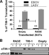
Expression of RAGE does not increase transduction. (A) Transduction of EBOV or LASV pseudovirions into HEK 293T cells transfected with an empty vector (Empty) or a RAGE-expressing vector (RAGE) relative to results with WT TIM-1. Cells were transduced at 48 h following transfection, and EGFP expression was assessed 24 h following transduction by flow cytometry. Data are shown as means ± SEM for at least three replicates. Significance compared to results for the WT was calculated using one-sample t-test comparison to 1 (**, P < 0.001). Significance between Empty and RAGE results was calculated using a two-sample t test. (B) TIM-1 and RAGE expression for three experiments, labeled 1 to 3, is shown by immunoblot analysis.
Fig 5
Fig 5
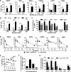
TIM-1-mediated transduction is not specific to EBOV. (A) Transduction of HEK 293T cells (gray bars) and TIM-1+ H3 cells (black bars) with VSVΔG pseudotyped with EBOV GP, RRV GP, GP64, CHIKV env, LASV GPC, or SINV 2.2 1L1L env. Cells were seeded in equal numbers, transduced with an MOI of ∼0.01 and 0.03, and assayed for EGFP by flow cytometry at 24 h following transduction. (B) Transduction of pseudovirions at an MOI of ∼0.01 into HEK 293T cells transfected with empty vector, ND115DN mutant of TIM-1, or WT TIM-1. MOIs were determined in untransfected HEK 293T cells. (C) Percent inhibition of pseudovirion transduction into TIM-1+ H3 cells by MAb ARD5, A6G2, or A8E5 (0.5 μg/ml). Percent inhibition is calculated relative to no-antibody control. (D) Transduction of pseudovirions into TIM-1+ H3 cells (filled) or HEK 293T cells (open) in the presence of 0, 5, or 25 μM PtdSer liposomes. (E) Correlation between enhancement of transduction by TIM-1 expression and percent inhibition by PtdSer liposomes. Pseudovirions were plotted on the x axis based on fold enhancement of transduction in H3 cells over that in HEK 293T cells and on the y axis based on percent inhibition by 25 μM PtdSer compared to that by 0 μM PtdSer liposomes. Linear regression was fitted and significance was calculated using the GraphPad Prism software program. (F) FIV pseudovirions expressing β-Gal were used to transduce HEK 293T cells and TIM-1+ H3 cells. β-Gal expression in lysates was assessed using the Galacto-Light system. Shown is the fold increase in β-Gal signal in H3 cells over that in HEK 293T cells. (G) Fold enhancement of VSVΔG pseudovirions transduction into HEK 293T cells transfected with TIM-1 or AnxVΔIgV-TIM-1 relative to that with the empty-vector control. Cells were transduced with an MOI of 0.01 as determined from titers in empty-vector HEK 293T cells. Data are shown as means ± SEM for at least three replicates. For panels A and B, significance was calculated using a two-sample student t test with equal variance, and for panels C and G, significance was calculated using one-sample t-test comparison to 0 (C) or 1 (G) (**, P < 0.001; *, P < 0.01).
Fig 6
Fig 6
Virus binding and internalization occur independent of glycoprotein. (A) Binding of FITC-labeled EBOV and No GP VSV pseudovirions to HEK 293T cells transfected with TIM-1, mutant TIM-1 ND115DN, or AnxVΔIgV-TIM-1 in the presence or absence of ARD5 (2 μg/ml). (B to E) Internalization of FITC-labeled EBOV and No GP pseudovirions (B and C) or VP40-GFP VLPs (D and E) into Vero cells. Virus was added at 4°C to Vero cells that were prebound with or without ARD5 (2 μg/ml) or PtdChl or PtdSer liposomes (25 μM). As noted, some cells were shifted to 37°C for 30 min, whereas others remained on ice. Identified cell populations were treated with trypsin and washed to remove excess virus. Fluorescence was determined by flow cytometry. Representative histograms are shown in panels A, B, and D, with a filled gray line representing background cellular fluorescence and a black line representing virus fluorescence. Histograms are quantified in panels C and E. Data are shown as means ± SD for at least three replicates.
Fig 7
Fig 7
RRV and AcMNPV infection is enhanced by TIM-1 expression. (A) Replication of RRV in TIM-1- or empty-vector-transfected cells at MOIs ranging from 0.001 to 0.1. Supernatants were collected 48 h following infection, and titers were determined by endpoint dilution on Vero cells. (B) Infection of TIM-1+ Vero cells with RRV in the presence or absence of MAb ARD5 (1 μg/ml). Supernatants were collected 48 h after infection and assessed for viral titer by endpoint dilution on Vero cells. (C) Transduction of HEK 293T cells or TIM-1+ H3 cells with recombinant baculovirus expressing β-Gal at an MOI of 0.003 as determined from titers on HEK 293T cells. After 48 h of incubation with virus, cells were lysed and β-Gal activity was assessed using the Galacto-Light system. (D) Impact of PtdSer on WT EBOV infection. Vero cells were preincubated for 1 h with the indicated concentrations of PtdSer or PtdChl liposomes or medium alone. Cells were then challenged in the presence of the liposomes with replication-competent EBOV encoding GFP. After 24 h, at which time the GFP from one round of infection can be detected, cells were fixed and the proportion of infected cells in the total cell population was determined (see Materials and Methods). The experiment was repeated 3 times with similar outcomes (50% inhibitory concentration [IC50], 5 ± 1.5 μM), and the means ± SD for 4 replicates are shown from 1 experiment. (E) Infection with mouse organ-derived EBOV GP-rVSV of Vero cells in the absence (black) or presence (gray) of ARD5. Virus obtained from lung, spleen, or kidney homogenates was serially diluted on Vero cells, and after 48 h, infection was assessed by EGFP expression. Data for RRV infections are shown as means ± SD, and AcMNPV and EBOV GP-rVSV data are shown as means ± SEM for at least three replicates. Significance was calculated using two-sample t-test comparison with equal variance (**, P < 0.001; *, P < 0.01).
Fig 8
Fig 8
Model for phosphatidylserine-mediated virus entry enhancing receptor (or PVEER) uptake of virus. (A) Virus buds from cell surface after infection, incorporating GP and PtdSer onto the viral envelope. The glycan cloud formed by extensive GP glycosylation events provides both stability of the GP in its prefusion state and potential steric protection against immune responses to GP residues. (B) Uptake of virus by a neighboring cell. PtdSer on the viral envelope interacts with PVEERs, such as the Gas6/Axl complex and TIM family, which mediate virus internalization. (C) Conditions within the endosome promote GP-dependent fusion events. Within the endosome, low-pH events can lead directly to GP conformational changes or, alternatively, protease processing of the GP, thereby reducing energy barriers required for fusion. Additional factors, such as binding of NPC1 by filovirus GPs, may also be necessary.

References

    1. Dolnik O, Kolesnikova L, Becker S. 2008. Filoviruses: interactions with the host cell. Cell. Mol. Life Sci. 65:756–776 - PMC - PubMed
    1. Geisbert TW, Young HA, Jahrling PB, Davis KJ, Larsen T, Kagan E, Hensley LE. 2003. Pathogenesis of Ebola hemorrhagic fever in primate models: evidence that hemorrhage is not a direct effect of virus-induced cytolysis of endothelial cells. Am. J. Pathol. 163:2371–2382 - PMC - PubMed
    1. Leroy EM, Gonzalez JP, Baize S. 2011. Ebola and Marburg haemorrhagic fever viruses: major scientific advances, but a relatively minor public health threat for Africa. Clin. Microbiol. Infect. 17:964–976 - PubMed
    1. Brindley MA, Hughes L, Ruiz A, McCray PB, Jr, Sanchez A, Sanders DA, Maury W. 2007. Ebola virus glycoprotein 1: identification of residues important for binding and postbinding events. J. Virol. 81:7702–7709 - PMC - PubMed
    1. Dube D, Brecher MB, Delos SE, Rose SC, Park EW, Schornberg KL, Kuhn JH, White JM. 2009. The primed ebolavirus glycoprotein (19-kilodalton GP1,2): sequence and residues critical for host cell binding. J. Virol. 83:2883–2891 - PMC - PubMed

Publication types