Review
. 2013 Jun;14(6):382-92.
doi: 10.1038/nrm3588.

Organization of the ER-Golgi interface for membrane traffic control

Affiliations
Review

Organization of the ER-Golgi interface for membrane traffic control

Federica Brandizzi et al. Nat Rev Mol Cell Biol. 2013 Jun.

Abstract

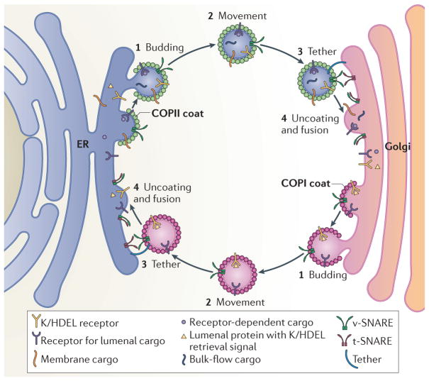
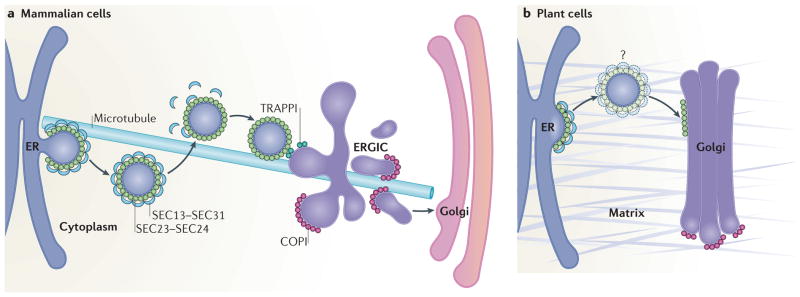
Coat protein complex I (COPI) and COPII are required for bidirectional membrane trafficking between the endoplasmic reticulum (ER) and the Golgi. While these core coat machineries and other transport factors are highly conserved across species, high-resolution imaging studies indicate that the organization of the ER-Golgi interface is varied in eukaryotic cells. Regulation of COPII assembly, in some cases to manage distinct cellular cargo, is emerging as one important component in determining this structure. Comparison of the ER-Golgi interface across different systems, particularly mammalian and plant cells, reveals fundamental elements and distinct organization of this interface. A better understanding of how these interfaces are regulated to meet varying cellular secretory demands should provide key insights into the mechanisms that control efficient trafficking of proteins and lipids through the secretory pathway.


Conflict of interest statement

Competing interests statement

The authors declare no competing financial interests.

Figures

Figure 1
Figure 1. Bidirectional transport between the ER and the Golgi is mediated by COPI and COPII carriers
Bidirectional transport of secretory cargo between the endoplasmic reticulum (ER) and the Golgi requires budding, movement, tethering, as well as uncoating and fusion of coat protein complex II (COPII) and COPI carriers with their respective compartments. These include bulk-flow cargo, membrane cargo and receptor-dependent luminal cargo. COPII carriers facilitate selective and bulk-flow cargo export towards the Golgi. One important function of COPI is to facilitate retrieval of escaped luminal proteins containing K/HDEL retrieval signals that are recognized by the K/HDEL receptor as well as other machinery required for optimal anterograde transport. Carrier fusion is mediated by vesicular SNARE proteins (v-SNAREs) and target-SNAREs (t-SNAREs) upon anchoring of the carriers to their target compartment via tethers.
Figure 2
Figure 2. The architecture of the COPII cage facilitates transport of diverse cargo
Structural analyses have demonstrated that the coat protein complex II (COPII) cage, which consists of the SEC13–SEC31 and SEC23–SEC24 subcomplexes, has a flexible architecture. a | Electron microscope reconstruction model of the SEC13–SEC31 cuboctahedral cage that is formed with purified mammalian SEC13–SEC31 (REF. 37) (Electron Microscopy Data Bank (EMDataBank) accession number: EMDB1232). b | Schematic representation of SEC31–SEC13 (shown in green and blue, respectively) heterotetramers arranged at a vertex point, indicating variable angles in the observed geometry of COPII cages. c | The structures for the SEC13–SEC31 assembly unit and the SEC13–SEC31 cage in the presence of SEC23 (REF. 147) and SEC23–SEC24 (REF. 35) have been reported. Variations in the SEC31 hinge (135°–165°) as well as in the β-vertex angle (90°–108°) of the SEC13–SEC31 cage have been documented and are listed in the table. The α-vertex angle is constant at 60° in each of the conditions. Under conditions in which SEC23–SEC24 is added to SEC13–SEC31, cuboctahedral (a 24 edged polygon) and icosidodecaheral (a 60 edged polygon) cage geometries were observed with each edge consisting of a SEC13–SEC31 heterotetramer. As the β-angle approaches 120° in the case of very large vesicles or tubules, the COPII cage may produce near-planar lattices. Distinct arrangements in the COPII cage are thought to allow for the range of COPII vesicle carriers necessary to accommodate varying cargo sizes.
Figure 3
Figure 3. The ER–Golgi interface and ERES have a distinct organization in mammals and plants
a | In mammalian cells, ER exit sites (ERES) are orientated towards a juxtaposed endoplasmic reticulum (ER)–Golgi intermediate compartment (ERGIC). Coat protein complex II (COPII)-coated vesicles originate within cup-shaped ER subdomains, which are associated with the plus end of microtubules. Upon fission of vesicles from the ERES, the SEC13–SEC31 cage is depolymerized, but the SEC23–SEC24 coat is partially retained. Vesicles reach the ERGIC in a microtubule-independent manner where they are tethered through the interaction between SEC23 and the TRAPPI (transport protein particle I) tethering complex. COPI mediates forward protein transport from the ERGIC towards the Golgi as well as recycling back to the ER membrane (the latter is not shown). b | In plant cells, ERES and Golgi are closely associated, possibly through a matrix (indicated in grey) that holds the ER and the Golgi together. The existence of COPII vesicles in plants is still debated (denoted by the question mark); vesicle-like structures have been seen,,, rarely in electron microscopy analyses of high-pressure frozen Arabidopsis thaliana tissues, although it was unclear whether they were undergoing budding or fusion. Unlike mammalian cells, plant cell ER–Golgi transport does not rely on the microtubule cytoskeleton.
Figure 4
Figure 4. Cellular architecture contributes to the ER–Golgi organization and positioning of ERES in mammalian and plant cells
a,b | Side-view schematic (a) or direct view (b) of a cultured mammalian cell, showing the relative positioning of the nucleus, endoplasmic reticulum (ER) network and the Golgi. The inset shows the distribution of ER exit sites (ERES), which are dispersed over the cytoplasmic ER network and concentrated in regions near the Golgi. c | Confocal image of hepatocarcinoma cells (HHU7 cells) co-transfected with SEC24–mCherry and VSVG–YFP, which label ERES and the ER, respectively. d | Side-view diagram of a plant cell showing that the ER is sandwiched between the tonoplast vacuole and the plasma membrane. e | Diagram illustrating the distribution of the ER network at the cortical region of a plant cell. The inset depicts the organization of the ER, ERES and closely associated Golgi complexes; the mobility of Golgi stacks is indicated by the dashed arrows. f | Confocal images of a tobacco leaf epidermal cell co-expressing the ER and Golgi marker ERD2–GFP (shown in green, right panel) and YFP–SEC24A (shown in red, middle panel). The merged image reveals a large overlap of fluorescent signals at punctae that represent Golgi and ERES. The image in panel c courtesy of K. Hirschberg, Tel Aviv University, Israel. The images in panel f courtesy of L. Renna, Michigan State University, USA.

Comment in

References

    1. Ward TH, Polishchuk RS, Caplan S, Hirschberg K, Lippincott-Schwartz J. Maintenance of Golgi structure and function depends on the integrity of ER export. J Cell Biol. 2001;155:557–570. - PMC - PubMed
    1. Brandizzi F, Snapp EL, Roberts AG, Lippincott-Schwartz J, Hawes C. Membrane protein transport between the endoplasmic reticulum and the Golgi in tobacco leaves is energy dependent but cytoskeleton independent: evidence from selective photobleaching. Plant Cell. 2002;14:1293–1309. - PMC - PubMed
    1. Altan-Bonnet N, Sougrat R, Lippincott-Schwartz J. Molecular basis for Golgi maintenance and biogenesis. Curr Opin Cell Biol. 2004;16:364–372. - PubMed
    1. Farquhar MG, Palade GE. The Golgi apparatus: 100 years of progress and controversy. Trends Cell Biol. 1998;8:2–10. - PMC - PubMed
    1. Driouich A, Staehelin LA. In: The Golgi Apparatus. Berger EG, Roth J, editors. Birkhäuser Verlag; 1997. pp. 275–301.

Publication types

Substances

LinkOut - more resources