. 2013 Jul 1;191(1):378-85.
doi: 10.4049/jimmunol.1300221. Epub 2013 May 22.

Low levels of insulin-like growth factor-1 contribute to alveolar macrophage dysfunction in cystic fibrosis

Affiliations

Low levels of insulin-like growth factor-1 contribute to alveolar macrophage dysfunction in cystic fibrosis

Jamie L Bessich et al. J Immunol. .

Abstract

Alveolar macrophages are major contributors to lung innate immunity. Although alveolar macrophages from cystic fibrosis (CF) transmembrane conductance regulator(-/-) mice have impaired function, no study has investigated primary alveolar macrophages in adults with CF. CF patients have low levels of insulin-like growth factor 1 (IGF-1), and our prior studies demonstrate a relationship between IGF-1 and macrophage function. We hypothesize that reduced IGF-1 in CF leads to impaired alveolar macrophage function and chronic infections. Serum and bronchoalveolar lavage (BAL) samples were obtained from eight CF subjects and eight healthy subjects. Macrophages were isolated from BAL fluid. We measured the ability of alveolar macrophages to kill Pseudomonas aeruginosa. Subsequently, macrophages were incubated with IGF-1 prior to inoculation with bacteria to determine the effect of IGF-1 on bacterial killing. We found a significant decrease in bacterial killing by CF alveolar macrophages compared with control subjects. CF subjects had lower serum and BAL IGF-1 levels compared with healthy control subjects. Exposure to IGF-1 enhanced alveolar macrophage macrophages in both groups. Finally, exposing healthy alveolar macrophages to CF BAL fluid decreased bacterial killing, and this was reversed by the addition of IGF-1, whereas IGF-1 blockade worsened bacterial killing. Our studies demonstrate that alveolar macrophage function is impaired in patients with CF. Reductions in IGF-1 levels in CF contribute to the impaired alveolar macrophage function. Exposure to IGF-1 ex vivo results in improved function of CF alveolar macrophages. Further studies are needed to determine whether alveolar macrophage function can be enhanced in vivo with IGF-1 treatment.


Figures
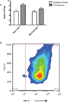
Figure 1
Figure 1. Isolation of alveolar macrophages from adult patients with cystic fibrosis
A. Subjects with CF underwent bronchoscopy with BAL. BAL fluid was filtered and neutrophils were depleted using magnetic beads for CD15. Total cell count was measured before and after neutrophil depletion and demonstrates similar macrophage numbers in CF subjects compared to healthy controls. B. Flow cytometry for the macrophage marker CD14 was performed on cells isolated from CF subjects. The image shown is representative of 3 separate experiments and demonstrates that 98.8% of cells isolated after neutrophil depletion are CD14+ macrophages.
Figure 2
Figure 2. CF alveolar macrophages upregulate the mannose receptor and down regulate TLR4
Alveolar macrophages were isolated from subjects with CF as described. Flow cytometry for the activated macrophage surface markers mannose receptor (MR) (panel A) and TLR4 (panel B) was performed on CF alveolar macrophages. The left panels demonstrate flow performed on unstained cells to determine the gating scheme. The images shown are representative of 3 separate experiments and demonstrate that 97.7% of cells are MR positive and 95.6% of cells are TLR4 negative.
Figure 3
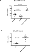
Figure 3. CF patients have reduced serum and BAL IGF-1 levels
A. Serum IGF-1 was measured by ELISA in CF subjects (n = 12) at baseline and during an acute exacerbation. IGF-1 was also measured in healthy controls (n = 8). ANOVA followed by Tukey’s test for multiple comparisons demonstrates significantly lower serum IGF-1 at baseline in CF subjects compared to healthy control (p<0.001). There was also a significant decrease in IGF-1 in CF subjects during an acute exacerbation compared to baseline (p<0.01). B. IGF-1 was measured in BAL fluid from CF subjects (n = 6) and healthy controls (n = 8). Student’s t-test demonstrates significantly lower IGF-1 levels in the CF subjects compared to healthy controls (p<0.001).
Figure 4
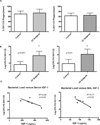
Figure 4. CF alveolar macrophages have impaired bacterial killing, which correlates with IGF-1 levels
A. Alveolar macrophages were isolated from CF subjects and healthy controls as described. Cells were exposed to the clinical P. aeruginosa strains DH1135 (left panel) and DH1133 (right panel) at an MOI of 10. After 1 hour, the supernatants were removed and the cells were lysed and the intracellular bacteria were quantified to determine the percentage of bacteria that were phagocytosed. Student’s t-test demonstrates no difference in the percentage of bacteria phagocytsoed by CF or healthy alveolar macrophages. B. Alveolar macrophages were isolated from CF subjects and healthy controls as described. Cells were exposed to the clinical P. aeruginosa strains DH1135 (left panel) and DH1133 (right panel) at an MOI of 10. After 1 hour, gentamicin (80 mg/mL) was added to the media to kill non-phagocytosed bacteria. After an additional 3 hours, remaining live bacteria were quantified. Student’s t-test demonstrates a significant decrease in bacterial killing by CF macrophages compared to healthy controls (p<0.001). Graph represents the mean and standard deviation of 4 separate experiments. C. IGF-1 was measured in serum and BAL fluid from the same CF subjects. Linear regression analysis demonstrates a significant inverse correlation between serum and BAL IGF-1 and remaining bacterial load (R2=0.58, p=0.05 for serum and R2=0.77, p=0.02 for BAL).
Figure 5
Figure 5. IGF-1 improves bacterial killing by healthy and CF alveolar macrophages
A. Alveolar macrophages were isolated from healthy volunteers as described. Cells were incubated for 40 hours with media and no IGF-1, low IGF-1 (100 ng/mL) or high IGF-1 (400 ng/mL). Cells were then exposed to P. aeruginosa DH1135. After 1 hour, gentamicin (80 mg/mL) was added to kill non-phagocytosed bacteria. After an additional 3 hours, remaining live bacteria were quantified. ANOVA followed by Tukey’s test for multiple comparisons demonstrates increased bacterial killing in the cells exposed to high IGF-1 compared to low IGF-1 (p=0.005) and no IGF-1 (p=0.002). Graph represents the mean and standard deviation of 4 separate experiments. B. Alveolar macrophages were isolated from CF subjects as described. Cells were incubated for 40 hours with media and no IGF-1, low IGF-1 (100ng/mL) or high IGF-1 (400ng/mL). Cells were then exposed to P. aeruginosa DH1135. After 1 hour, gentamicin (80 mg/mL) was added to kill non-phagocytosed bacteria. After an additional 3 hours, remaining live bacteria were quantified. ANOVA followed by Tukey’s test for multiple comparisons demonstrates increased bacterial killing in the cells exposed to high IGF-1 compared to low IGF-1 (p=0.014) and no IGF-1 (p=0.0062). Graph represents the mean and standard deviation of 4 separate experiments.
Figure 6
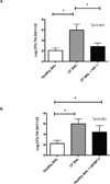
Figure 6. CF BAL fluid causes impaired killing by healthy alveolar macrophages that is improved by IGF-1
Alveolar macrophages were isolated from healthy volunteers as described. A. Cells were exposed to 10% healthy BAL fluid, 10% CF BAL fluid, or 10% CF BAL fluid plus 400 ng/mL IGF-1 for 40 hours. Cells were then exposed to P. aeruginosa DH1135. After 1 hour, gentamicin (80 mg/mL) was added to kill non-phagocytosed bacteria. After an additional 3 hours, remaining live bacteria were quantified. ANOVA followed by Tukey’s test for multiple comparisons demonstrates decreased bacterial killing in the cells exposed to CF BAL fluid compared to healthy BAL fluid (p<0.001). There was also a significant improvement in bacterial killing in cells treated with CF BAL fluid and IGF-1 compared to CF BAL fluid alone (p<0.001). B. Cells were exposed to 10% healthy BAL fluid, 10% CF BAL fluid, or 10% healthy BAL fluid plus IGFBP-2 (2µg/mL). Bacterial killing was assessed as described above and demonstrated decreased bacterial killing after addition of IGF1BP-2 (p<0.001). Graphs represent the mean and standard deviation of 3 separate experiments.

References

    1. Rowe SM, Miller S, Sorscher EJ. Cystic fibrosis. N Engl J Med. 2005;352:1992–2001. - PubMed
    1. Deriy LV, Gomez EA, Zhang G, Beacham DW, Hopson JA, Gallan AJ, Shevchenko PD, Bindokas VP, Nelson DJ. Disease-causing mutations in the cystic fibrosis transmembrane conductance regulator determine the functional responses of alveolar macrophages. J Biol Chem. 2009;284:35926–35938. - PMC - PubMed
    1. Di A, Brown ME, Deriy LV, Li C, Szeto FL, Chen Y, Huang P, Tong J, Naren AP, Bindokas V, et al. CFTR regulates phagosome acidification in macrophages and alters bactericidal activity. Nat Cell Biol. 2006;8:933–944. - PubMed
    1. Stoltz DA, Meyerholz DK, Pezzulo AA, Ramachandran S, Rogan MP, Davis GJ, Hanfland RA, Wohlford-Lenane C, Dohrn CL, Bartlett JA, et al. Cystic fibrosis pigs develop lung disease and exhibit defective bacterial eradication at birth. Sci Transl Med. 2010;2 29ra31. - PMC - PubMed
    1. Del Porto P, Cifani N, Guarnieri S, Di Domenico EG, Mariggio MA, Spadaro F, Guglietta S, Anile M, Venuta F, Quattrucci S, et al. Dysfunctional CFTR alters the bactericidal activity of human macrophages against Pseudomonas aeruginosa. PLoS One. 2011;6:e19970. - PMC - PubMed

Publication types

MeSH terms

Substances