Review
. 2013 Jun;77(2):267-76.
doi: 10.1128/MMBR.00003-13.

Pyrophosphate-fueled Na+ and H+ transport in prokaryotes

Affiliations
Review

Pyrophosphate-fueled Na+ and H+ transport in prokaryotes

Alexander A Baykov et al. Microbiol Mol Biol Rev. 2013 Jun.

Erratum in

  • Microbiol Mol Biol Rev. 2013 Sep;77(3):540

Abstract

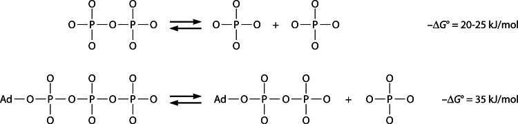
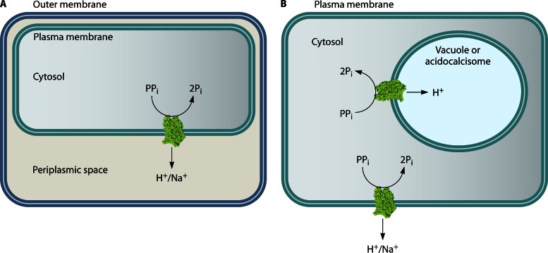
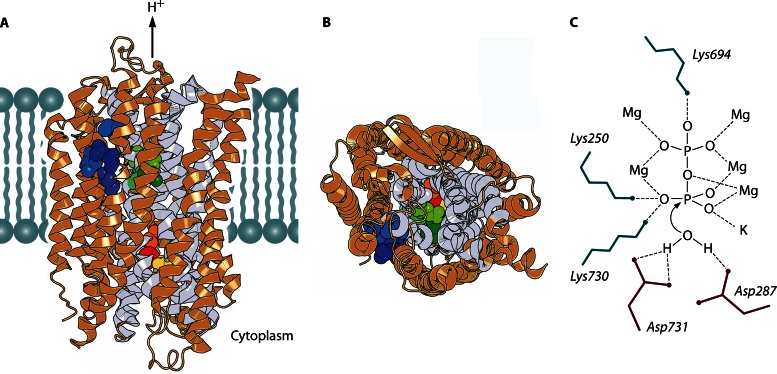
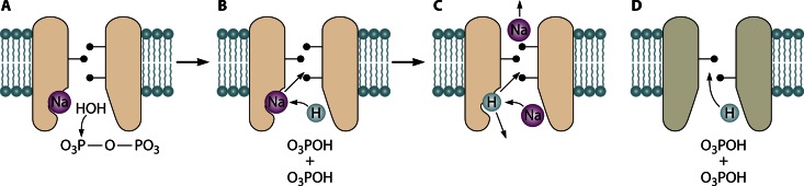
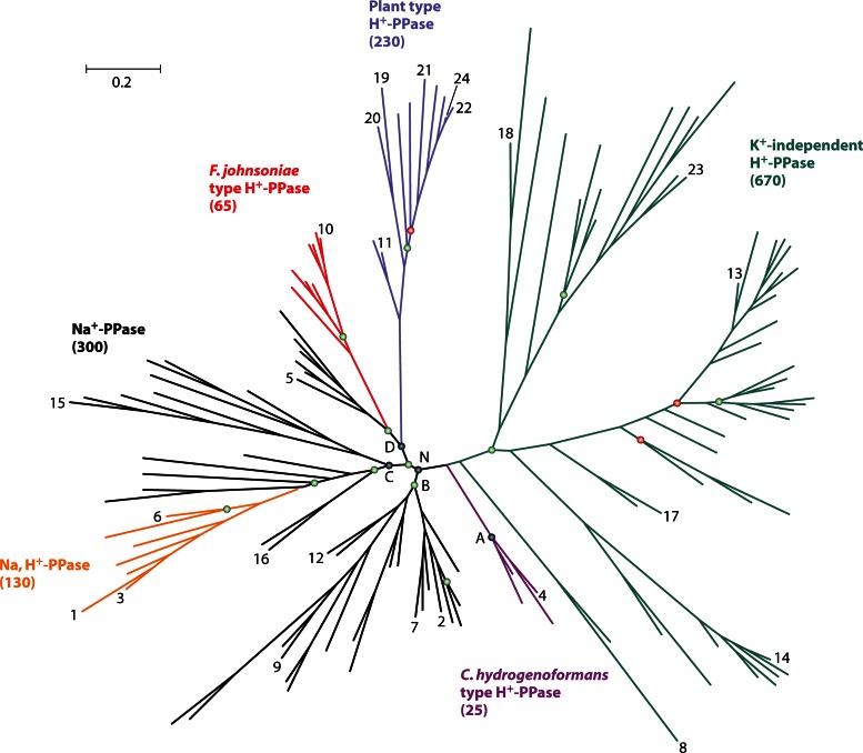
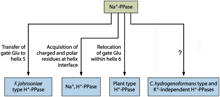
In its early history, life appeared to depend on pyrophosphate rather than ATP as the source of energy. Ancient membrane pyrophosphatases that couple pyrophosphate hydrolysis to active H(+) transport across biological membranes (H(+)-pyrophosphatases) have long been known in prokaryotes, plants, and protists. Recent studies have identified two evolutionarily related and widespread prokaryotic relics that can pump Na(+) (Na(+)-pyrophosphatase) or both Na(+) and H(+) (Na(+),H(+)-pyrophosphatase). Both these transporters require Na(+) for pyrophosphate hydrolysis and are further activated by K(+). The determination of the three-dimensional structures of H(+)- and Na(+)-pyrophosphatases has been another recent breakthrough in the studies of these cation pumps. Structural and functional studies have highlighted the major determinants of the cation specificities of membrane pyrophosphatases and their potential use in constructing transgenic stress-resistant organisms.


Figures

Fig 1
Fig 1
Hydrolyses of PPi to Pi and of ATP to ADP and Pi yield appreciable energy that can be used to drive coupled chemical or transport reactions. Ad, adenosine moiety.
Fig 2
Fig 2
Orientation of membrane PPase and ion fluxes in prokaryotic (A) and eukaryotic (B) cells. Acidocalcisomes are acidic phosphorus and cation storage organelles that were originally identified in protists (7) and resemble plant vacuoles. Acidocalcisomes with membrane-bound PPases are also present in some bacteria (8, 9). To date, only H+-translocating PPases have been identified in acidocalcisomes and vacuoles. In both cases, the PPase active site faces the cytosol, and cations are transported out of it. H+-PPases localized to the plasma membrane and Golgi apparatus have also been reported for plant cells.
Fig 3
Fig 3
Structure of V. radiata H+-PPase (Protein Data Bank [PDB] accession number 4A01) (11) and presumed coordination of the substrate. (A and B) Two views of the PPase subunit (in the plane of and perpendicular to the membrane) showing bound imidodiphosphate (yellow), K+ ion (red), residues occupying the position that determines K+ dependence (A537) (orange), alternative positions of the gate-forming residue (E301/G297/S235) (shades of green), and positions specific for Na+,H+-PPase (L148/T152/V204/I227) (shades of blue). Imidodiphosphate, K+, and the indicated residues are shown as spheres; the remainder of the molecule is presented as a ribbon diagram. The inner-circle helices are shown in gray, and the rest of the molecule is shown in light brown. The right side of the subunit in panels A and B forms a contact with the second subunit. (C) Presumed coordination of PPi and the water nucleophile in the active site. Ionic interactions and hydrogen bonds are depicted as dashed lines. Six of the nine PPi ligands are protein-bound metal ions (Mg2+ and K+). PPi hydrolysis proceeds via direct attack of the water molecule without formation of a phosphorylated intermediate. Panels A and B were created with PyMOL (PyMOL Molecular Graphics System, version 1.5.0.4; Schrödinger, LLC).
Fig 4
Fig 4
Possible elements of the coupling mechanisms in membrane PPases. (A to C) Three key events in the “billiards-type” mechanism of Na+-transporting PPases: Na+, PPi, and nucleophilic water binding (A); PPi hydrolysis and displacement of Na+ from its binding site into the ion conductance channel by the H+ ion generated from the nucleophilic water (B); and subsequent displacement of the bound H+ ion by incoming Na+ (C). Depending on whether the H+ ion is released into the medium or the ion conductance channel, the transporter is classified as Na+-PPase or Na+,H+-PPase, respectively. (D) Direct entrance of the H+ ion generated during PPi hydrolysis into the ion conductance channel in H+-PPases, which lack the transitional binding site for Na+ and H+.
Fig 5
Fig 5
Simplified phylogenetic tree of verified and putative membrane PPases. The sequences in the tree were selected by redundancy filtering to leave only representative sequences for each group of highly similar sequences. The total number of sequences found in the NCBI protein sequence database for each PPase subfamily is given in parentheses. The tree consists of one K+-independent and five K+-dependent subfamilies, shown in different colors. K+-independent H+-PPases occur in bacteria, archaea, plants, and protists; C. hydrogenoformans-type H+-PPases, F. johnsoniae-type H+-PPases, and Na+,H+-PPases occur in bacteria only; plant-type H+-PPases occur in plants, protists, and bacteria (Leptospira); and Na+-PPases occur in bacteria and archaea (a few protists and algae may also have Na+-PPase genes). The letters A, B, C, D, and N indicate the nodes of different clades within the K+-dependent family. The nodes with MrBayes clade credibility values in the ranges of 70 to 84 and 50 to 69 are marked with green and red circles, respectively; for all other nodes, the credibility values were 85 to 100. The scale bar represents 0.2 substitutions per residue. The PPases with experimentally verified cation specificity are indicated as follows: 1, Akkermansia muciniphila; 2, Anaerostipes caccae; 3, B. vulgatus; 4, C. hydrogenoformans; 5, Chlorobium limicola; 6, Clostridium leptum; 7, C. tetani; 8, C. thermocellum; 9, Desulfuromonas acetoxidans; 10, F. johnsoniae; 11, Leptospira biflexa; 12, M. thermoacetica; 13, R. rubrum; 14, S. coelicolor; and 15, T. maritima (bacteria); 16, M. mazei (Na+-PPase); 17, M. mazei (H+-PPase); and 18, Pyrobaculum aerophilum (archaea); 19, Plasmodium falciparum; 20, Toxoplasma gondii; and 21, Trypanosoma cruzi (protists); 22, Arabidopsis thaliana (AVP1); 23, Arabidopsis thaliana (AVP2); and 24, V. radiata (plants).
Fig 6
Fig 6
Four presumed routes from Na+ transport to H+ transport in membrane PPase.

Similar articles

Cited by

References

    1. Baltscheffsky H. 1996. Energy conversion leading to the origin and early evolution of life: did inorganic pyrophosphate precede adenosine triphosphate?, p 1–9 In Baltscheffsky H. (ed), Origin and evolution of biological energy conversion. VCH, New York, NY
    1. Holm NG, Baltscheffsky H. 2011. Links between hydrothermal environments, pyrophosphate, Na+, and early evolution. Orig. Life Evol. Biosph. 41:483–493 - PMC - PubMed
    1. Heinonen JK. 2001. Biological role of inorganic pyrophosphate. Kluwer Academic Publishers, London, United Kingdom
    1. Seufferheld MJ, Kim KM, Whitfield J, Valerio A, Caetano-Anollés G. 2011. Evolution of vacuolar proton pyrophosphatase domains and volutin granules: clues into the early evolutionary origin of the acidocalcisome. Biol. Direct 6:50 doi:10.1186/1745-6150-6-50 - DOI - PMC - PubMed
    1. Baltscheffsky H, Von Stedingk LV, Heldt HW, Klingenberg M. 1966. Inorganic pyrophosphate: formation in bacterial photophosphorylation. Science 153:1120–1122 - PubMed

Publication types

LinkOut - more resources