. 2013 Jul;24(14):2216-27.
doi: 10.1091/mbc.E12-12-0883. Epub 2013 May 22.

PakB binds to the SH3 domain of Dictyostelium Abp1 and regulates its effects on cell polarity and early development

Affiliations

PakB binds to the SH3 domain of Dictyostelium Abp1 and regulates its effects on cell polarity and early development

Yidai Yang et al. Mol Biol Cell. 2013 Jul.

Abstract

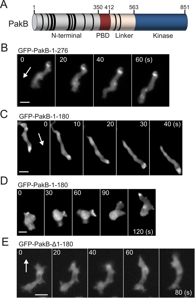
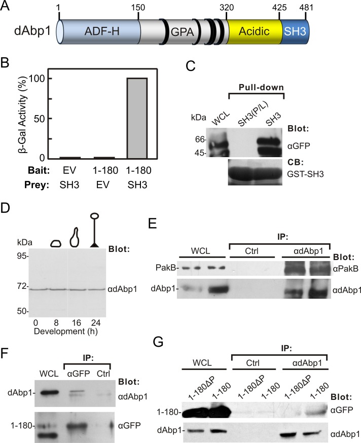
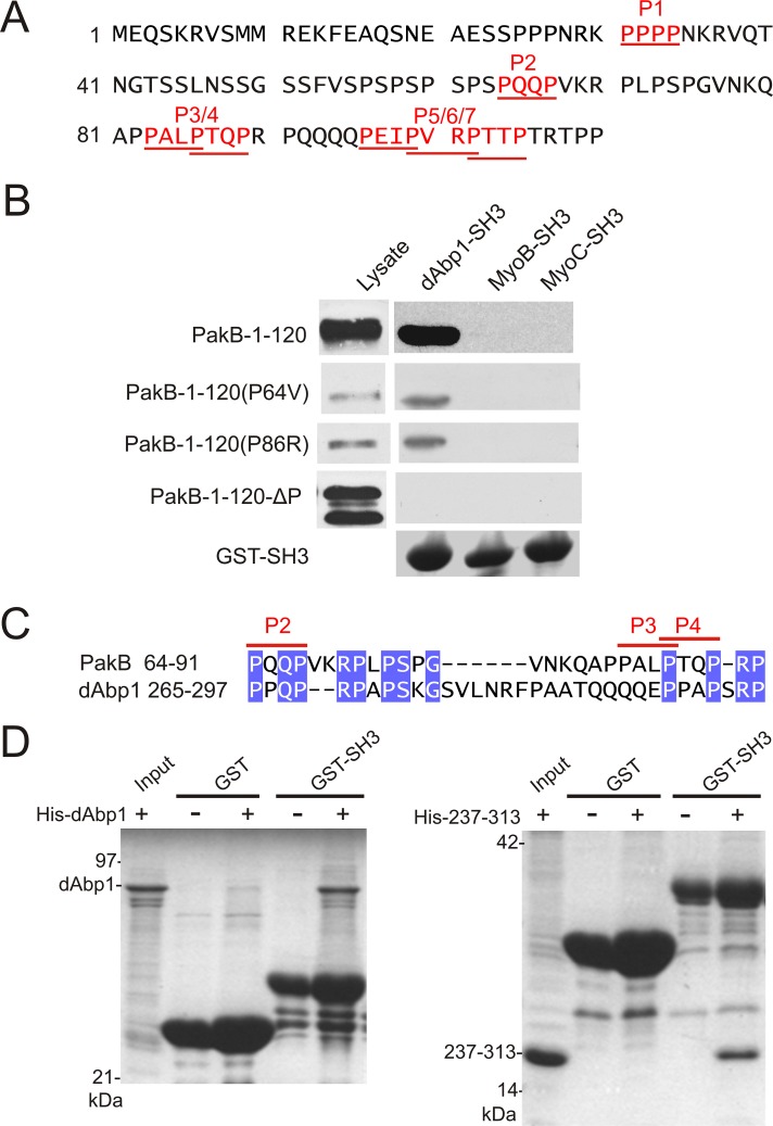
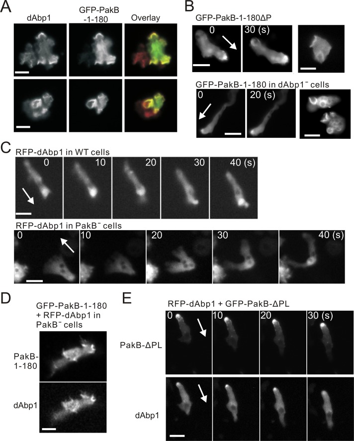
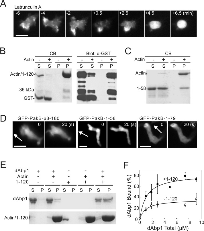
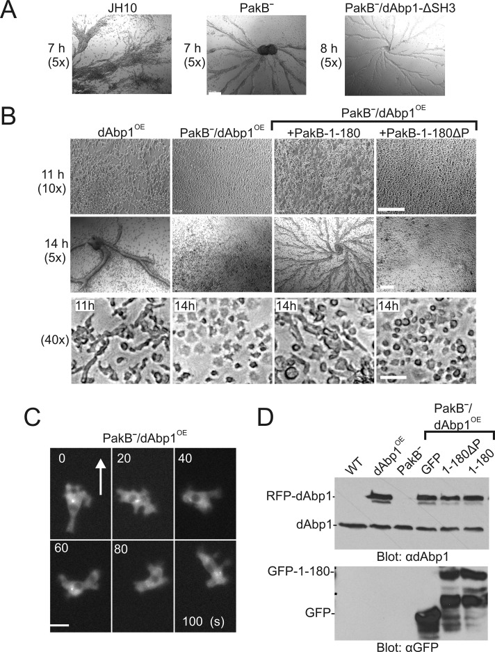
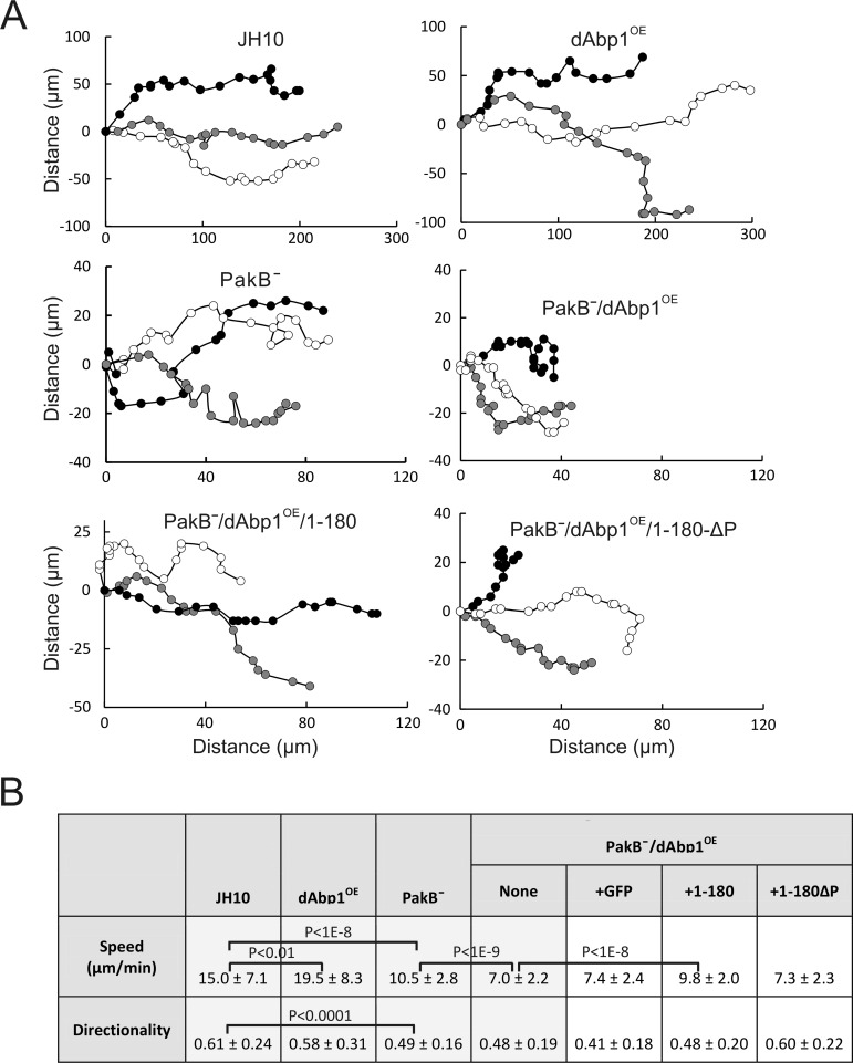
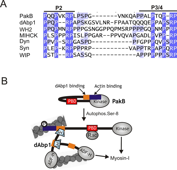
Dictyostelium p21-activated kinase B (PakB) phosphorylates and activates class I myosins. PakB colocalizes with myosin I to actin-rich regions of the cell, including macropinocytic and phagocytic cups and the leading edge of migrating cells. Here we show that residues 1-180 mediate the cellular localization of PakB. Yeast two-hybrid and pull-down experiments identify two proline-rich motifs in PakB-1-180 that directly interact with the SH3 domain of Dictyostelium actin-binding protein 1 (dAbp1). dAbp1 colocalizes with PakB to actin-rich regions in the cell. The loss of dAbp1 does not affect the cellular distribution of PakB, whereas the loss of PakB causes dAbp1 to adopt a diffuse cytosolic distribution. Cosedimentation studies show that the N-terminal region of PakB (residues 1-70) binds directly to actin filaments, whereas dAbp1 exhibits only a low affinity for filamentous actin. PakB-1-180 significantly enhances the binding of dAbp1 to actin filaments. When overexpressed in PakB-null cells, dAbp1 completely blocks early development at the aggregation stage, prevents cell polarization, and significantly reduces chemotaxis rates. The inhibitory effects are abrogated by the introduction of a function-blocking mutation into the dAbp1 SH3 domain. We conclude that PakB plays a critical role in regulating the cellular functions of dAbp1, which are mediated largely by its SH3 domain.


Figures

FIGURE 1:
FIGURE 1:
PakB-1-180 mimics the subcellular localization of PakB. (A) Dictyostelium PakB consists of a proline-rich, N-terminal domain, a p21-binding domain (PBD) that recognizes Rac GTPases, an unstructured linker segment, and a C-terminal Ser/Thr protein kinase domain. The positions of PxxP motifs are indicated by black rings. (B) Time-course images of GFP-PakB-1-276 expressed in an aggregation-competent AX3 cell migrating in a cAMP gradient. (C, D) Time-course images of GFP-PakB-1-180 expressed in (C) an aggregation-competent AX3 cell migrating in a cAMP gradient and (D) a growth-phase AX3 cell extending pseudopods and macropinocytic cups. (E) Time-course images of GFP-PakB lacking residues 1–180 (GFP-PakB-Δ1-180) expressed in an AX3 cell. Arrows indicate the direction of migration. Bars, 10 μm.
FIGURE 2:
FIGURE 2:
PakB-1-180 binds the dAbp1 SH3 domain. (A) dAbp1 consists of an ADF-H domain, a segment rich in glutamine, proline, and alanine (GPA) residues, a highly acidic region, and an SH3 domain. The positions of PxxP motifs are indicated by black rings. (B) Two-hybrid analysis was carried out using a bait vector expressing PakB-1-180 (1–180) and a prey vector expressing the dAbp1 SH3 domain (SH3) and empty bait or prey vectors (EV). The strength of the interaction was assessed quantitatively by liquid culture β-galactosidase assay. (C) Pull-down assays were carried out using the GST-dAbp1-SH3 domain (SH3) or an inactive GST-dAbp1-SH3 domain (P474L mutation; SH3(P/L)) and lysates of cells expressing GFP-PakB-1-180. The whole-cell lysate (WCL) and washed pellets were probed using an anti-GFP antibody. Coomassie blue (CB) was used to visualize the GST-SH3 domains in the pellets. (D) Immunoblot analysis of AX3 cells harvested at different stages of development using an affinity-purified rabbit polyclonal antibody to dAbp1. (E) Coimmunoprecipitation of endogenous dAbp1 and PakB. Immunoprecipitates were prepared from AX3 cell lysates using a control (Ctrl) or anti-dAbp1 antibody. The WCL and washed immunoprecipitates were immunoblotted using anti-PakB and anti-dAbp1 antibodies. Results from two experiments are shown. (F) Immunoprecipitates were prepared from lysates of cells expressing GFP-PakB-1-180 (1–180) using an anti-GFP antibody or a control (Ctrl) antibody. The WCL and washed immunoprecipitates were immunoblotted using antibodies to GFP and dAbp1. (G) Immunoprecipitates were prepared from cells expressing GFP-PakB-1-180 (1–180) or GFP-PakB-1-180ΔP (1-180ΔP) using an anti-dAbp1 or a control (Ctrl) antibody. The WCLs and washed immunoprecipitates were immunoblotted using antibodies to GFP and dAbp1.
FIGURE 3:
FIGURE 3:
Identification of two binding sites for the dAbp1 SH3 domain in PakB. (A) The N-terminal region of PakB contains seven PxxP motifs (P1–P7; red underlined). (B) Pull-down assays were performed using GST fusion proteins containing the SH3 domains of dAbp1, MyoB, or MyoC and lysates of PakBˉ cells expressing the indicated GFP-PakB constructs. PakB-1-120 containing both the P64V and P86 mutations is designated PakB-1-120-ΔP. The cell lysates and washed pellets were subjected to immunoblot analysis using an anti-GFP antibody. GST-SH3 domains were visualized by Coomassie blue staining. (C) dAbp1 contains a proline-rich sequence similar to the P2–P3/4 region of PakB. (D) Pull-down assays were performed using GST (GST) or GST-dAbp1-SH3 (GST-SH3) and His-tagged dAbp1(His-dAbp1; left) or residues 237–313 of dAbp1 (His-237-313; right). The washed pellets were visualized by Coomassie blue staining after SDS–PAGE.
FIGURE 4:
FIGURE 4:
Colocalization of PakB and dAbp1. (A) AX3 cells expressing GFP-PakB-1-180 were fixed and stained using antibodies against dAbp1 and GFP. The overlay shows staining for GFP in green and dAbp1 in red. (B) Images of GFP-PakB-1-180ΔP expressed in PakBˉ cells (top) and GFP-PakB-1-180 expressed in dAbp1-null (dAbp1) cells (bottom). Migrating developed cells are shown in the left and middle, and growth-phase cells with macropinocytic cups are shown in the right. (C) RFP-dAbp1 was imaged in a developed migrating AX3 cell (top) and PakBˉ cell (bottom). (D) Expression of GFP-PakB-1-180 in a PakBˉ cell restores the cortical localization of RFP-dAbp1. (E) Expression of constitutively active GFP-PakB-ΔPL, which mislocalizes to the rear of migrating cells, results in recruitment of RFP-dAbp1 to the cell posterior. Arrows indicate the direction of migration. Bars, 10 μm.
FIGURE 5:
FIGURE 5:
The PakB N-terminus binds actin. (A) Time-course images showing the redistribution of PakB-GFP to the cytoplasm after addition of latrunculin A (5 μM) at time 0. Bar, 10 μm. (B, C) Actin filament cosedimentation experiments were performed using (B) GST-PakB-1-120 (1–120) and (C) GST-PakB-1-58 (1–58). Samples were centrifuged at 100,000 × g for 30 min, and the resulting supernatants (S) and pellets (P) were subjected to SDS–PAGE and stained with Coomassie blue (B, left; C) or probed with an anti-GST antibody (B, right). Note that GST-PakB-1-120 and actin have the same mobility on SDS–PAGE. (D) Images of developed migrating PakBˉ cells expressing GFP-PakB-1-58, GFP-PakB-1-79, or GFP-PakB-68-180. Arrows indicate the direction of migration. Bars, 10 μm. (E) Actin filament cosedimentation experiments were performed with GST-dAbp1 in the presence or absence of GST-PakB-1-120 (1–120). Samples were centrifuged at 100,000 × g for 30 min, and the resulting supernatants (S) and pellets (P) were subjected to SDS–PAGE and stained with Coomassie blue. (F) The percentage of GST-dAbp1 that cosedimented with actin in the presence (·) or absence (◯) of 5 μM GST-PakB-1-120 was quantified by densitometry of Coomassie blue–stained SDS gels. Results show the mean and SD for three independent experiments.
FIGURE 6:
FIGURE 6:
Overexpression of dAbp1 blocks development of PakBˉ cells. (A) JH10 cells, PakBˉ cells, and PakBˉ cells expressing RFP-dAbp1 with a function-blocking mutation in its SH3 domain (dAbp1-ΔSH3) were starved to induce development. The cells formed streams and aggregates after 7-8 h of starvation. Magnification, 5×. (B) JH10 cells expressing RFP-dAbp1 (dAbp1OE), PakBˉ cells expressing RFP-dAbp1 (PakBˉ/dAbp1OE), and PakBˉ/dAbp1OE cells expressing GFP-PakB-1-180 or GFP-PakB-1-180ΔP were starved to induce development and imaged at 7, 11, or 14 h at 5, 10, or 40× magnification as indicated. Bar, 0.2 mm (top, middle), 20 μm (bottom). (C) Time-course images of a developed PakBˉ/dAbp1OE cell migrating in a cAMP gradient. The cell undergoes rapid shape changes, with RFP-dAbp1 diffusely distributed in the cytoplasm. Arrow indicates the direction of migration. Bar, 10 μm. (D) Extracts of the described cell lines were subject to immunoblot analysis using anti-dAbp1 (top) and anti-GFP (bottom) antibodies to show the expression levels of dAbp1, RFP-dAbp1, GFP-PakB-1-180, and PakB-1-180ΔP.
FIGURE 7:
FIGURE 7:
Overexpression of dAbp1 in PakBˉ cells inhibits chemotaxis. (A) Developed cells were monitored during chemotaxis in a linear and stable cAMP gradient using Ibidi μ-slides. Positions were recorded every 30 s. Typical tracks for three cells from each of the indicated cell lines are shown. (B) Speed and directionality values were calculated for each cell line using the Ibidi Chemotaxis and Migration Tool 2.0. Results show the mean and standard deviations for 15–20 cells in each of three separate experiments. Cells were tracked for a total of 20 min.
FIGURE 8:
FIGURE 8:
PakB interacts with the dAbp1 SH3 domain via a conserved proline-rich sequence. (A) Sequences similar to the two binding sites for the dAbp1 SH3 domain in PakB (P2 and P3/4; residues 64–91) are conserved in dAbp1 (residues 265–297), a Dictyostelium WH2 domain–containing protein (WH2; residues 361-396), Acanthamoeba myosin I heavy chain kinase (MIHCK; residues 207–239), human dynamin-1 (Dyn; residues 813–836); human synaptojanin-1 (Syn; residues 1141–1165), and human WAS/WASL-interacting protein family member 1 (WIP-1; residues 295–316). GenBank accession numbers are as follows: WH2 domain-containing protein (EAL66623), MIHCK (AAD09141), dynamin-1 (AAH50279), synaptojanin-1 (NP_003886), and WIP-1 (NP_003378). (B) A model for the PakB–dAbp1 complex. Inactive PakB is believed to adopt a folded conformation in which the N-terminal region suppresses the activity of the PBD and kinase domains. Autophosphorylation of Ser-8, which occurs in the absence of allosteric effectors, allows GTP-Rac to bind the PBD domain (red box) and increases kinase activity 40-fold. It is proposed that Ser-8 autophosphorylation causes PakB to unfold, which would increase exposure of the N-terminal region that contains an actin-binding module (blue box) and binding sites for the dAbp1 SH3 domain (orange box). The actin-binding module recruits PakB to actin-rich regions at the cell cortex, where it serves as a docking site for dAbp1. Whether the P2 and P3/4 motifs can support the concurrent binding of two dAbp1 molecules is not known. dAbp1 contains an ADF-H domain that weakly binds actin filaments and a P2-P3/4-like sequence (orange) that binds its own SH3 domain, suggesting that dimers or oligomers of dAbp1 may assemble to cross-link and strengthen the local actin network. dAbp1 interacts directly with MyoK, and possibly other class I myosins, suggesting that it may facilitate their recruitment to the complex to be phosphorylated and activated by PakB.

Similar articles

Cited by

References

    1. Bement WM, Mooseker MS. TEDS rule: a molecular rationale for differential regulation of myosins by phosphorylation of the heavy chain head. Cell Motil Cytoskeleton. 1995;31:87–92. - PubMed
    1. Brzeska H, Young R, Knaus U, Korn ED. Myosin I heavy chain kinase: cloning of the full-length gene and acidic lipid-dependent activation by rac and cdc42. Proc Natl Acad Sci USA. 1999;96:394–399. - PMC - PubMed
    1. Chen CL, Wang Y, Sesaki H, Iijima M. Myosin I links PIP3 signaling to remodeling of the actin cytoskeleton in chemotaxis. Sci Signal. 2012;5:ra10. - PMC - PubMed
    1. Cortesio CL, Perrin BJ, Bennin DA, Huttenlocher A. Actin-binding protein-1 interacts with WASp-interacting protein to regulate growth factor-induced dorsal ruffle formation. Mol Biol Cell. 2010;21:186–197. - PMC - PubMed
    1. Dai J, Ting-Beall HP, Hochmuth RM, Sheetz MP, Titus MA. Myosin I contributes to the generation of resting cortical tension. Biophys J. 1999;77:1168–1176. - PMC - PubMed

Publication types

MeSH terms

LinkOut - more resources