

Accumulation of abnormal adult-generated hippocampal granule cells predicts seizure frequency and severity
- PMID: 23699504
- PMCID: PMC3731053
- DOI: 10.1523/JNEUROSCI.5161-12.2013
Accumulation of abnormal adult-generated hippocampal granule cells predicts seizure frequency and severity
Abstract
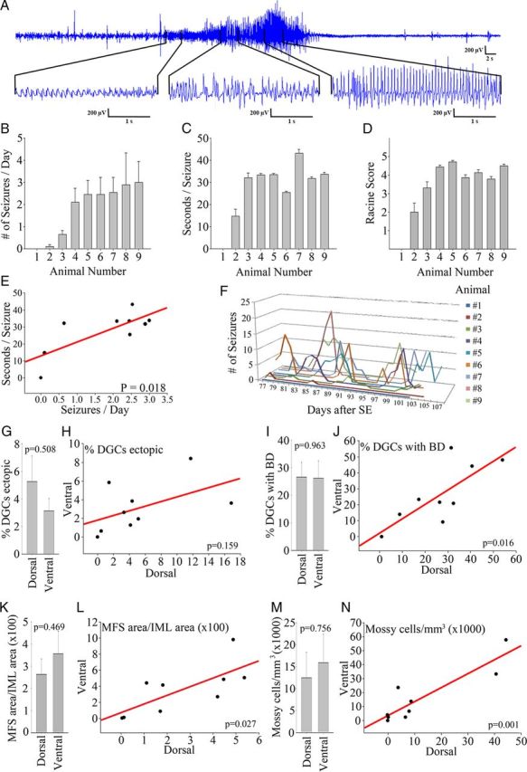
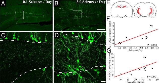
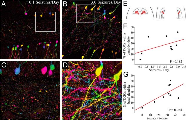
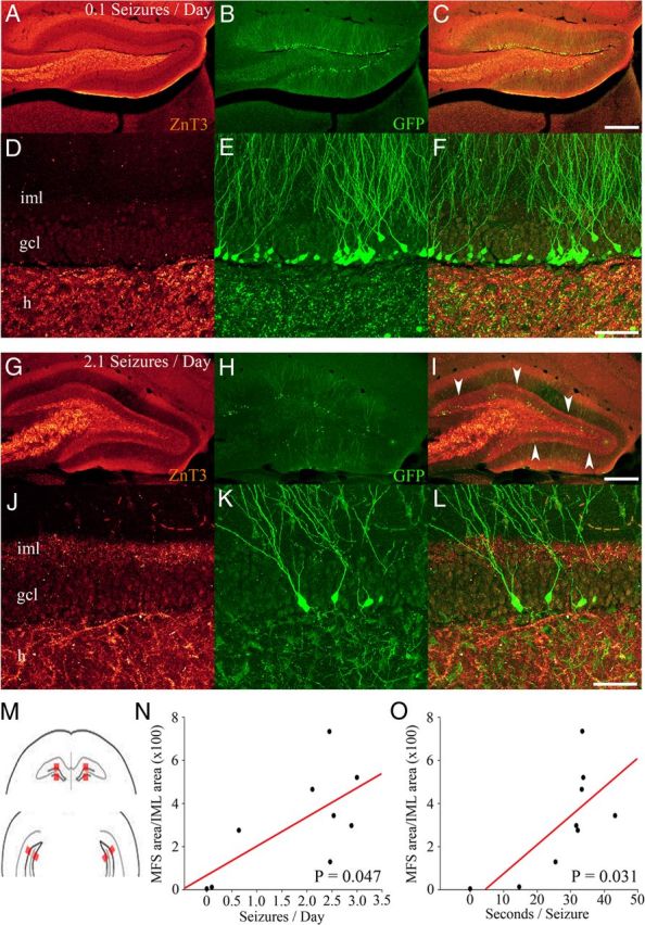
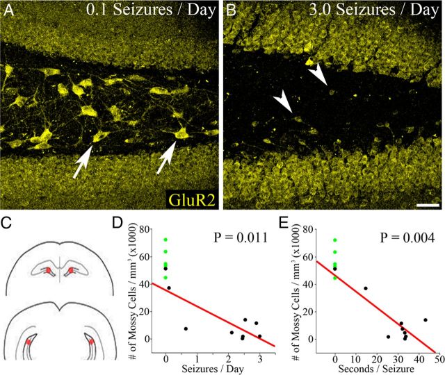
Accumulation of abnormally integrated, adult-born, hippocampal dentate granule cells (DGCs) is hypothesized to contribute to the development of temporal lobe epilepsy (TLE). DGCs have long been implicated in TLE, because they regulate excitatory signaling through the hippocampus and exhibit neuroplastic changes during epileptogenesis. Furthermore, DGCs are unusual in that they are continually generated throughout life, with aberrant integration of new cells underlying the majority of restructuring in the dentate during epileptogenesis. Although it is known that these abnormal networks promote abnormal neuronal firing and hyperexcitability, it has yet to be established whether they directly contribute to seizure generation. If abnormal DGCs do contribute, a reasonable prediction would be that the severity of epilepsy will be correlated with the number or load of abnormal DGCs. To test this prediction, we used a conditional, inducible transgenic mouse model to fate map adult-generated DGCs. Mossy cell loss, also implicated in epileptogenesis, was assessed as well. Transgenic mice rendered epileptic using the pilocarpine-status epilepticus model of epilepsy were monitored continuously by video/EEG for 4 weeks to determine seizure frequency and severity. Positive correlations were found between seizure frequency and (1) the percentage of hilar ectopic DGCs, (2) the amount of mossy fiber sprouting, and (3) the extent of mossy cell death. In addition, mossy fiber sprouting and mossy cell death were correlated with seizure severity. These studies provide correlative evidence in support of the hypothesis that abnormal DGCs contribute to the development of TLE and also support a role for mossy cell loss.
Figures
Similar articles
-
Impact of rapamycin on status epilepticus induced hippocampal pathology and weight gain.Exp Neurol. 2016 Jun;280:1-12. doi: 10.1016/j.expneurol.2016.03.015. Epub 2016 Mar 17. Exp Neurol. 2016. PMID: 26995324 Free PMC article.
-
Abnormalities of granule cell dendritic structure are a prominent feature of the intrahippocampal kainic acid model of epilepsy despite reduced postinjury neurogenesis.Epilepsia. 2012 May;53(5):908-21. doi: 10.1111/j.1528-1167.2012.03463.x. Epilepsia. 2012. PMID: 22533643 Free PMC article.
-
Excessive activation of mTOR in postnatally generated granule cells is sufficient to cause epilepsy.Neuron. 2012 Sep 20;75(6):1022-34. doi: 10.1016/j.neuron.2012.08.002. Neuron. 2012. PMID: 22998871 Free PMC article.
-
Chronic epileptogenic cellular alterations in the limbic system after status epilepticus.Epilepsia. 1999;40 Suppl 1:S23-33; discussion S40-1. doi: 10.1111/j.1528-1157.1999.tb00875.x. Epilepsia. 1999. PMID: 10421558 Review.
-
Temporal Lobe Epileptogenesis: A Focus on Etiology, Neuron Loss, the Latent Period, and Dentate Granule Cell Disinhibition.In: Noebels JL, Avoli M, Rogawski MA, Vezzani A, Delgado-Escueta AV, editors. Jasper's Basic Mechanisms of the Epilepsies. 5th edition. New York: Oxford University Press; 2024. Chapter 24. In: Noebels JL, Avoli M, Rogawski MA, Vezzani A, Delgado-Escueta AV, editors. Jasper's Basic Mechanisms of the Epilepsies. 5th edition. New York: Oxford University Press; 2024. Chapter 24. PMID: 39637110 Free Books & Documents. Review.
Cited by
-
In vivo reprogramming for tissue repair.Nat Cell Biol. 2015 Mar;17(3):204-11. doi: 10.1038/ncb3108. Nat Cell Biol. 2015. PMID: 25720960 Review.
-
Enriched Environment Altered Aberrant Hippocampal Neurogenesis and Improved Long-Term Consequences After Temporal Lobe Epilepsy in Adult Rats.J Mol Neurosci. 2015 Jun;56(2):409-21. doi: 10.1007/s12031-015-0571-0. Epub 2015 May 7. J Mol Neurosci. 2015. PMID: 25946980
-
Normal and epilepsy-associated pathologic function of the dentate gyrus.Prog Brain Res. 2016;226:155-78. doi: 10.1016/bs.pbr.2016.04.005. Epub 2016 May 18. Prog Brain Res. 2016. PMID: 27323942 Free PMC article. Review.
-
Gq-Coupled Muscarinic Receptor Enhancement of KCNQ2/3 Channels and Activation of TRPC Channels in Multimodal Control of Excitability in Dentate Gyrus Granule Cells.J Neurosci. 2019 Feb 27;39(9):1566-1587. doi: 10.1523/JNEUROSCI.1781-18.2018. Epub 2018 Dec 28. J Neurosci. 2019. PMID: 30593498 Free PMC article.
-
Electroacupuncture at ST36-ST37 and at ear ameliorates hippocampal mossy fiber sprouting in kainic acid-induced epileptic seizure rats.Biomed Res Int. 2014;2014:756019. doi: 10.1155/2014/756019. Epub 2014 Jun 22. Biomed Res Int. 2014. PMID: 25045697 Free PMC article.
References
Publication types
MeSH terms
Substances
Grants and funding
LinkOut - more resources
Full Text Sources
Other Literature Sources
Medical
Molecular Biology Databases