. 2013 May 22;33(21):9104-12.
doi: 10.1523/JNEUROSCI.0299-13.2013.

Neuronal mechanisms of respiratory pattern generation are evolutionary conserved

Affiliations

Neuronal mechanisms of respiratory pattern generation are evolutionary conserved

Elenia Cinelli et al. J Neurosci. .

Abstract

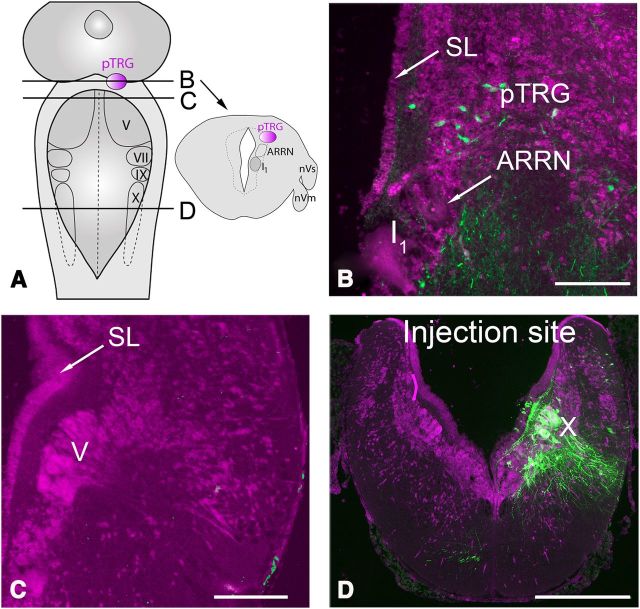
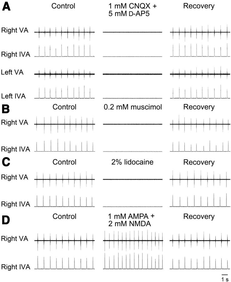
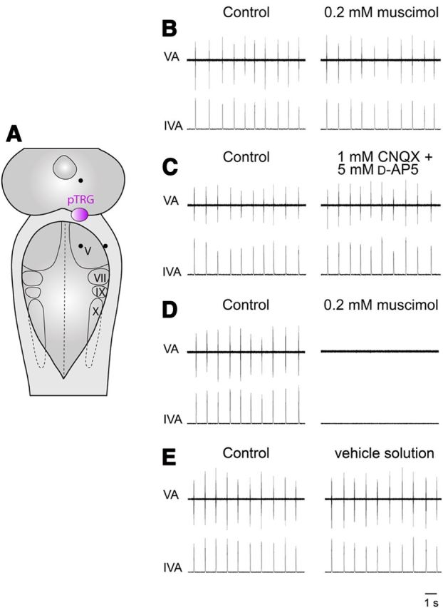
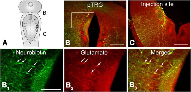
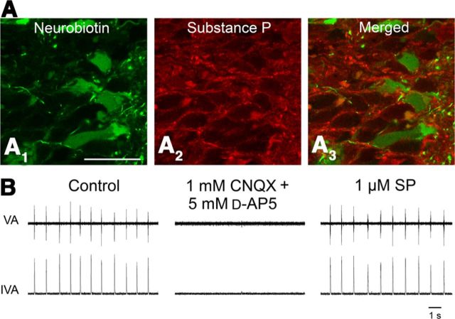
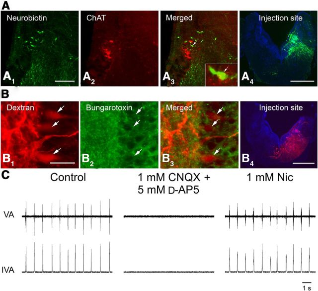
A brainstem region, the paratrigeminal respiratory group (pTRG), has been suggested to play a crucial role in the respiratory rhythm generation in lampreys. However, a detailed characterization of the pTRG region is lacking. The present study performed on isolated brainstem preparations of adult lampreys provides a more precise localization of the pTRG region with regard to both connectivity and neurochemical markers. pTRG neurons projecting to the vagal motoneuronal pool were identified in a restricted area of the rostral rhombencephalon at the level of the isthmic Müller cell I1 close to sulcus limitans of His. Unilateral microinjections of lidocaine, muscimol, or glutamate antagonists into the pTRG inhibited completely the bilateral respiratory activity. In contrast, microinjections of glutamate agonists enhanced the respiratory activity, suggesting that this region is critical for the respiratory pattern generation. The retrogradely labeled pTRG neurons are glutamatergic and surrounded by terminals with intense substance P immunoreactivity. Cholinergic neurons were seen close to, and intermingled with, pTRG neurons. In addition, α-bungarotoxin binding sites (indicating nicotinic receptors) were found throughout the pTRG area and particularly on the soma of these neurons. During apnea, induced by blockade of ionotropic glutamate receptors within the same region, microinjections of 1 μm substance P or 1 mm nicotine into the pTRG restored rhythmic respiratory activity. The results emphasize the close similarities between the pTRG and the mammalian pre-Bötzinger complex as a crucial site for respiratory rhythmogenesis. We conclude that some basic features of the excitatory neurons proposed to generate respiratory rhythms are conserved throughout evolution.


Figures

Figure 1.
Figure 1.
Distribution of retrogradely labeled neurons in the pTRG region of the lamprey. A, Schematic illustration of a dorsal view of the lamprey mesencephalon/rhombencephalon showing the levels of the coronal sections illustrated in B–D (unbroken lines) and the location of the pTRG (pink area). The pTRG location is also reported (pink area) on a schematic coronal section (arrow). B, Photomicrograph of a transverse section of the isthmic region showing retrogradely labeled neurons (green signal) after injections of Neurobiotin into the vagal motoneuron pool. A population of labeled cells was found at the level of the anterior rhomboencephalic reticular nucleus close to the isthmic Müller cell I1, in a region corresponding to the pTRG. C, Section at the level of the trigeminal motor nucleus ∼60 μm caudal to the section reported in B showing the absence of retrogradely labeled cells after tracer injections into the vagal motoneuron pool. D, Photomicrograph of a transverse section of the caudal rhomboencephalon showing the site of Neurobiotin injection into the vagal motor nucleus (green signal). Note the small bundle of crossing fibers. All sections are counterstained with deep red fluorescent Nissl stain. I1, Isthmic Müller cell; nVm, motor root of the trigeminal nerve; nVs, sensory root of the trigeminal nerve; SL, sulcus limitans of His; V, trigeminal motor nucleus; VII, facial motor nucleus; IX, glossopharyngeal motor nucleus; X, vagal motor nucleus. Scale bars: B, C, 200 μm; D, 500 μm.
Figure 2.
Figure 2.
Examples of respiratory responses evoked by blockade or activation of pTRG region. A, Suppression of respiratory rhythmic activity ∼1 min after unilateral microinjections into the pTRG region of a mixture of 1 mm CNQX and 5 mm d-AP5. B, C, Similar effects on respiration induced by unilateral microinjections into the pTRG region of 0.2 mm muscimol or 2% lidocaine, respectively. D, Increases in respiratory frequency and peak vagal activity ∼2 min after a unilateral microinjection of a mixture of 1 mm AMPA and 2 mm NMDA into the pTRG. VA, Raw vagal nerve activity; IVA, integrated vagal nerve activity.
Figure 3.
Figure 3.
Unilateral control microinjections. A, Control injection sites (●) 0.4 mm or more away from the pTRG (pink area) shown on a schematic illustration of a dorsal view of the lamprey mesencephalon/rhombencephalon. B, Absence of appreciable respiratory effects ∼1 min after a unilateral microinjection of 0.2 mm muscimol at the lateral border of the trigeminal motor nucleus. C, Absence of obvious respiratory responses ∼1 min after a unilateral microinjection of a mixture of 1 mm CNQX and 5 mm d-AP5 into the mesencephalic region. The drugs used failed to induce the characteristic effects elicited by unilateral microinjections into the pTRG in all the selected control sites. D, The dramatic inhibitory effects on respiratory activity induced by a unilateral microinjection of 0.2 mm muscimol into the pTRG have also been illustrated for comparison. E, Absence of obvious respiratory responses ∼1 min after a unilateral microinjection of the vehicle solution into the pTRG. V, Trigeminal motor nucleus; VII, facial motor nucleus; IX, glossopharyngeal motor nucleus; X, vagal motor nucleus; VA, raw vagal nerve activity; IVA, integrated vagal nerve activity.
Figure 4.
Figure 4.
Distribution of glutamate immunoreactivity in the pTRG. A, Schematic illustration of a dorsal view of the lamprey mesencephalon/rhombencephalon showing the levels of the coronal sections reported in B and C (unbroken lines). B, Photomicrograph of a transverse section at the level of the isthmic area/rostral rhombencephalon showing retrogradely labeled neurons (merged, Neurobiotin green plus glutamate immunoreactivity red signals) in the pTRG region after injections of Neurobiotin into the vagal motoneuron pool. Labeled fibers can be observed crossing at the isthmic roof. C, Transverse section of the caudal rhombencephalon showing the site of Neurobiotin injection (merged, Neurobiotin green plus glutamate immunoreactivity red signals) into the vagal nucleus. B1–B3, Photomicrographs at a higher magnification of the portion of the transverse section indicated by the white rectangle in B, showing retrogradely labeled neurons (B1), glutamate immunoreactivity (B2), and merged image (B3) at the level of the pTRG region. Retrogradely labeled neurons displaying immunoreactivity for glutamate are indicated by white arrows. V, Trigeminal motor nucleus; VII, facial motor nucleus; IX, glossopharyngeal motor nucleus; X, vagal motor nucleus. Scale bars: B, C, 200 μm; B1–B3, 100 μm.
Figure 5.
Figure 5.
Rhythmogenic role of substance P. A, Substance P immunoreactivity within the pTRG. Confocal photomicrographs showing retrogradely labeled neurons (A1, green signal) after injections of Neurobiotin into the vagal motoneuron pool, substance P immunoreactivity (A2, red signal), and merged image (A3). Note the presence within the pTRG region of some retrogradely labeled neurons surrounded by substance P-immunoreactive structures. Scale bar: A1–A3, 30 μm. B, Bilateral microinjections of 1 mm CNQX/5 mm d-AP5 into the pTRG abolished the respiratory rhythm that was restored ∼1 min after bilateral microinjections of 1 μm substance P (SP) into the same site. VA, Raw vagal nerve activity; IVA, integrated vagal nerve activity.
Figure 6.
Figure 6.
Rhythmogenic role of acetylcholine. A, Immunofluorescent labeling of ChAT-expressing neurons in the pTRG area. A1, Photomicrographs of retrogradely labeled neurons (green signal) after injections of Neurobiotin into the region of vagal motoneurons. A2, ChAT immunostaining (red signal). A3, Merged image. Note the presence of a small population of cholinergic cells grouped close to retrogradely labeled neurons. The inset (6× magnification) shows ChAT-immunoreactive dots in close apposition to a retrogradely labeled cell (white arrows). A4, Photomicrograph of a transverse section of the caudal rhomboencephalon showing the site of Neurobiotin injections into the vagal motoneuronal pool (green signal). Cells are counterstained with blue fluorescent Nissl stain. B, Distribution of α-bungarotoxin binding sites in the pTRG area. B1, Photomicrographs of retrogradely labeled neurons (red signal) after injections of Texas Red-conjugated dextran into the region of vagal motoneurons. B2, α-Bungarotoxin binding sites (green signal). B3, Merged image. Retrogradely labeled neurons (white arrows in B1–B3) are surrounded by α-bungarotoxin binding sites. B4, Photomicrograph of a transverse section of the caudal rhombencephalon showing the injection site of Texas Red-conjugated dextran into the vagal motoneuronal pool (red signal). Cells are counterstained with blue fluorescent Nissl stain. Scale bars: A1–A3, 200 μm; A4, B4, 500 μm; B1–B3, 25 μm. C, Bilateral microinjections of 1 mm CNQX/5 mm d-AP5 into the pTRG abolished the respiratory rhythm that was restored ∼1 min after bilateral microinjections of 1 mm nicotine (Nic) into the same sites. VA, Raw vagal nerve activity; IVA, integrated vagal nerve activity.

Similar articles

Cited by

References

    1. Albuquerque EX, Pereira EF, Alkondon M, Rogers SW. Mammalian nicotinic acetylcholine receptors: from structure to function. Physiol Rev. 2009;89:73–120. doi: 10.1152/physrev.00015.2008. - DOI - PMC - PubMed
    1. Auclair F, Lund JP, Dubuc R. Immunohistochemical distribution of tachykinins in the CNS of the lamprey Petromyzon marinus. J Comp Neurol. 2004;479:328–346. doi: 10.1002/cne.20324. - DOI - PubMed
    1. Ben-Mabrouk F, Tryba AK. Substance P modulation of TRPC3/7 channels improves respiratory rhythm regularity and ICAN-dependent pacemaker activity. Eur J Neurosci. 2010;31:1219–1232. doi: 10.1111/j.1460-9568.2010.07156.x. - DOI - PMC - PubMed
    1. Ben-Mabrouk F, Amos LB, Tryba AK. Metabotropic glutamate receptors (mGluR5) activate transient receptor potential canonical channels to improve the regularity of the respiratory rhythm generated by the pre-Botzinger complex in mice. Eur J Neurosci. 2012;35:1725–1737. doi: 10.1111/j.1460-9568.2012.08091.x. - DOI - PMC - PubMed
    1. Bongianni F, Deliagina TG, Grillner S. Role of glutamate receptor subtypes in the lamprey respiratory network. Brain Res. 1999;826:298–302. doi: 10.1016/S0006-8993(99)01251-2. - DOI - PubMed

Publication types

MeSH terms

LinkOut - more resources