. 2013 May 22;33(21):9113-21.
doi: 10.1523/JNEUROSCI.5043-12.2013.

Protection from noise-induced hearing loss by Kv2.2 potassium currents in the central medial olivocochlear system

Affiliations

Protection from noise-induced hearing loss by Kv2.2 potassium currents in the central medial olivocochlear system

Huaxia Tong et al. J Neurosci. .

Abstract

The central auditory brainstem provides an efferent projection known as the medial olivocochlear (MOC) system, which regulates the cochlear amplifier and mediates protection on exposure to loud sound. It arises from neurons of the ventral nucleus of the trapezoid body (VNTB), so control of neuronal excitability in this pathway has profound effects on hearing. The VNTB and the medial nucleus of the trapezoid body are the only sites of expression for the Kv2.2 voltage-gated potassium channel in the auditory brainstem, consistent with a specialized function of these channels. In the absence of unambiguous antagonists, we used recombinant and transgenic methods to examine how Kv2.2 contributes to MOC efferent function. Viral gene transfer of dominant-negative Kv2.2 in wild-type mice suppressed outward K(+) currents, increasing action potential (AP) half-width and reducing repetitive firing. Similarly, VNTB neurons from Kv2.2 knock-out mice (Kv2.2KO) also showed increased AP duration. Control experiments established that Kv2.2 was not expressed in the cochlea, so any changes in auditory function in the Kv2.2KO mouse must be of central origin. Further, in vivo recordings of auditory brainstem responses revealed that these Kv2.2KO mice were more susceptible to noise-induced hearing loss. We conclude that Kv2.2 regulates neuronal excitability in these brainstem nuclei by maintaining short APs and enhancing high-frequency firing. This safeguards efferent MOC firing during high-intensity sounds and is crucial in the mediation of protection after auditory overexposure.


Figures
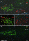
Figure 1.
Figure 1.
Kv2.2 is exclusively expressed in the MNTB and VNTB nuclei of the SOC. a, Coimmunostaining of the MNTB, VNTB, and the superior paraolivary nucleus (SPN) with antibodies to Kv3.1b (green) and Kv2.2 (red); nuclei are stained with DAPI (blue). MNTB principal neurons also show intense labeling for Kv3.1b (green), whereas the VNTB exhibits only Kv2.2 staining. b, Kv2.2 is present in MNTB neurons, but is restricted to the axon initial segment and is particularly intense in the medial part of the nucleus. c, The smaller VNTB cells show somatic staining for Kv2.2. d, Tissue from a Kv2.2KO mouse shows similar Kv3.1 labeling in the MNTB but no labeling with Kv2.2 antibodies in either MNTB or VNTB. Scale bars, 50 μm. Top is dorsal and left is medial.
Figure 2.
Figure 2.
Kv2.2 enables high-frequency firing. a, Outward K+ currents from WT-MNTB neurons (black) are much larger than those from a Kv2.2KO mouse (red). Recordings were made within 20 min of the killing of the animal. Voltage protocol is shown below. b, I/V relationship for average K+ currents measured in the MNTB show large magnitude currents WT (black) compared with the smaller currents from the Kv2.2KO (red). c, In current-clamp, a 400 Hz train of short depolarizing stimuli reliably induced APs in response to every stimulus repetition in WT-MNTB, whereas neurons from the Kv2.2KO exhibited failures (arrows). d, On average, WT MNTB neurons reliably followed stimulus trains up to 1000 Hz, with entrainment never dropping below 80%. Entrainment in Kv2.2KO neurons dropped significantly for stimulation frequencies of 400 Hz or above. e, Outward K+ currents in WT-VNTB neurons (black) were large and slightly inactivating, whereas K+ currents in Kv2.2KO VNTB neurons were strongly inactivating (reflecting the absence of slowly activating Kv2.2 currents (KO, red). f, Steady-state I/Vs were significantly different for VNTB neurons between genotypes. g, VNTB neurons are less able to follow high-frequency stimulus trains even in WT mice (black), but Kv2.2KO VNTB neurons cannot even follow a 100 Hz stimulus train without skipping cycles (red). h, The average ability to follow stimulation in VNTB neurons during repetitive stimulation shows high transmission at frequencies up to 200 Hz in the WT, but responses only to every other cycle in the Kv2.2KO.
Figure 3.
Figure 3.
Kv2.2 is not expressed in the cochlea. a, b, WT (a) and Kv2.2KO (b) brainstem sections (transverse plane) served as positive and negative controls, respectively, confirming that the Kv2.2 antibody is specific under the modified protocol required for cochlea sectioning and immunohistochemistry. c, Immunostaining of organ of Corti cross-sections in the cochlea from WT mice showed Kv2.1 staining, but no specific Kv2.2 immunoreactivity was observed. d, Identical staining was observed in the Kv2.2KO tissue. Open arrows indicate OHCs; filled arrow, IHCs. Representative images from WT (n = 3) and KO (n = 3) are shown. Scale bar, 20 μm.
Figure 4.
Figure 4.
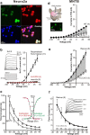
Suppression and rescue of Kv2.2 currents by respective transfection with viral vectors carrying Kv2.2DN or Kv2.2WT. a, Confocal imaging shows neuro2a cells transfected with Kv2.2WT virus. Green EGFP of transfected cells are stained by a Kv2.2 antibody (red, Kv2.2); cell nuclei are indicated by DAPI (blue) and the overlay is shown (bottom right). b, Whole-cell voltage-clamp shows large recombinant Kv2.2 currents from EGFP-positive cells after Kv2.2WT rAd infection (inset traces) with the mean I/V for 11 cells (black triangle). No currents were evoked in noninfected control cells (black circle) or when infected with the Kv2.2DN virus (open triangle). c, Recombinant Kv2.2WT activation rates were fit with a single exponential and are highly voltage dependent: the mean activation time constant is plotted against voltage and shows an e-fold acceleration with 18.4 mV depolarization (n = 11) at room temperature (black triangle). Faster activation rates were observed at 32°C (gray open triangle, e-fold acceleration with 17.2 mV depolarization, n = 9). Inset shows activation (green) and inactivation (red) curves for the recombinant Kv2.2WT currents; fits to Boltzmann functions gave half-activation of −9.5 mV and half-inactivation was at −38.8 mV. d, I/V relation for MNTB neurons in the presence of TEA (1 mm, filled circle) and after Kv2.2DN rAd expression (open circle), shows clear suppression of native K+ currents from injected animals. The inset shows a schematic of unilateral in vivo rAd vector injection to the mouse MNTB (top) and EGFP-positive MNTB neurons (bottom) from an injected mouse killed 4 d later. e, TEA-insensitive K+ currents are much greater in Kv2.2WT vector-injected Kv2.2KO mice (Rescue, filled square) than those from control Kv2.2KO mice (open square). f, In the presence of TEA, MNTB neurons from rescued mice (filled square) exhibited classic Kv2.2 activation kinetics that were much slower than those from Kv2.2KO mice (open square). The insert shows representative current traces from a rescued (black) and a Kv2.2KO (gray) MNTB neuron at indicated voltages. Both current traces were normalized to the same amplitude to highlight the slower activation of the Kv2.2 current. n is indicated in brackets.
Figure 5.
Figure 5.
Kv2.2 channels are essential for maintaining AP firing patterns in both MNTB (left) and VNTB (right) neurons. a, b, Lack of functional Kv2.2 channels prolonged AP half-width in Kv2.2KO (red) and Kv2.2DN (blue) compared with WT (black) for neurons from both MNTB (a) and VNTB (b). Bar graphs show average data for the change in AP half-width in the MNTB (aii) and in the VNTB (bii); n values are given by the number in the bars. c, Example voltage traces (ci) in response to 400 pA current injection, showed the characteristic single AP resonse in a WT MNTB neuron (black trace), but multiple APs were generated in Kv2.2KO mice (red trace) or after Kv2.2DN (blue trace). Neurons expressing Kv2.2DN or from Kv2.2KO mice fired trains of APs in contrast to the single AP firing phenotype of WT. The average number of APs evoked during a 200 ms current step is plotted against the injected current magnitude (cii). d, Example traces (di) show that VNTB AP firing is maintained throughout a step current injection (WT, black trace), whereas AP firing in the Kv2.2KO (red trace) and the Kv2.2DN (blue trace) fails to be maintained. The plot of mean AP firing (number of APs in 200 ms, dii) against injected current shows that the Kv2.2KO and Kv2.2DN VNTB neurons cannot sustain firing during large depolarizations.
Figure 6.
Figure 6.
Kv2.2 channels in the MOC/VNTB neurons enhance protection from AOE. a, Cross-section of the auditory brainstem shows afferent pathways (red) from the cochlea to the SOC, via the ventral cochlear nucleus (VCN). The MNTB and VNTB are shown in yellow. The VNTB neurons provide part of the MOC efferent projection (green) to the cochlea; the MNTB gives an inhibitory projection (blue) to the other nuclei of the SOC. The inset image shows dextran-rhodamine (red)-labeled VNTB neurons, which were transported from the cochlea, and with the same cells also immunolabeled with Kv2.2 (green). b, ABRs measured in vivo had similar thresholds in naive WT and Kv2.2KO mice across a frequency range of 8–30 kHz. c, Representative examples of ABRs measured in vivo in response to click stimulation. WT mice (black) and Kv2.2KO (red) mice show up to four clearly distinguishable components in the ABR traces (labeled I-IV) and no genotype-specific difference in waveforms or threshold was observed. d, Acoustic insult diminished all ABR components in the Kv2.2KO (red), indicating that the Kv2.2KO is much more sensitive to an acoustic insult, whereas WT animals (black) showed considerable recovery 1 week after the insult. Bar graph shows the significantly elevated mean ABR thresholds (*p < 0.05) in Kv2.2KO mice 1 week after inducing an acoustic insult.

References

    1. Ashmore J. Cochlear outer hair cell motility. Physiol Rev. 2008;88:173–210. doi: 10.1152/physrev.00044.2006. - DOI - PubMed
    1. Ballestero J, Zorrilla de San Martín J, Goutman J, Elgoyhen AB, Fuchs PA, Katz E. Short-term synaptic plasticity regulates the level of olivocochlear inhibition to auditory hair cells. J Neurosci. 2011;31:14763–14774. doi: 10.1523/JNEUROSCI.6788-10.2011. - DOI - PMC - PubMed
    1. Barnes-Davies M, Forsythe ID. Pre- and postsynaptic glutamate receptors at a giant excitatory synapse in rat auditory brainstem slices. J Physiol. 1995;488:387–406. - PMC - PubMed
    1. Boyev KP, Liberman MC, Brown MC. Effects of anesthesia on efferent-mediated adaptation of the DPOAE. J Assoc Res Otolaryngol. 2002;3:362–373. doi: 10.1007/s101620020044. - DOI - PMC - PubMed
    1. Brew HM, Forsythe ID. Two voltage-dependent K+ conductances with complementary functions in postsynaptic integration at a central auditory synapse. J Neurosci. 1995;15:8011–8022. - PMC - PubMed

Publication types

MeSH terms

LinkOut - more resources