. 2013 May 22;33(21):9169-75.
doi: 10.1523/JNEUROSCI.0301-13.2013.

The SNARE proteins SNAP25 and synaptobrevin are involved in endocytosis at hippocampal synapses

Affiliations

The SNARE proteins SNAP25 and synaptobrevin are involved in endocytosis at hippocampal synapses

Zhen Zhang et al. J Neurosci. .

Abstract

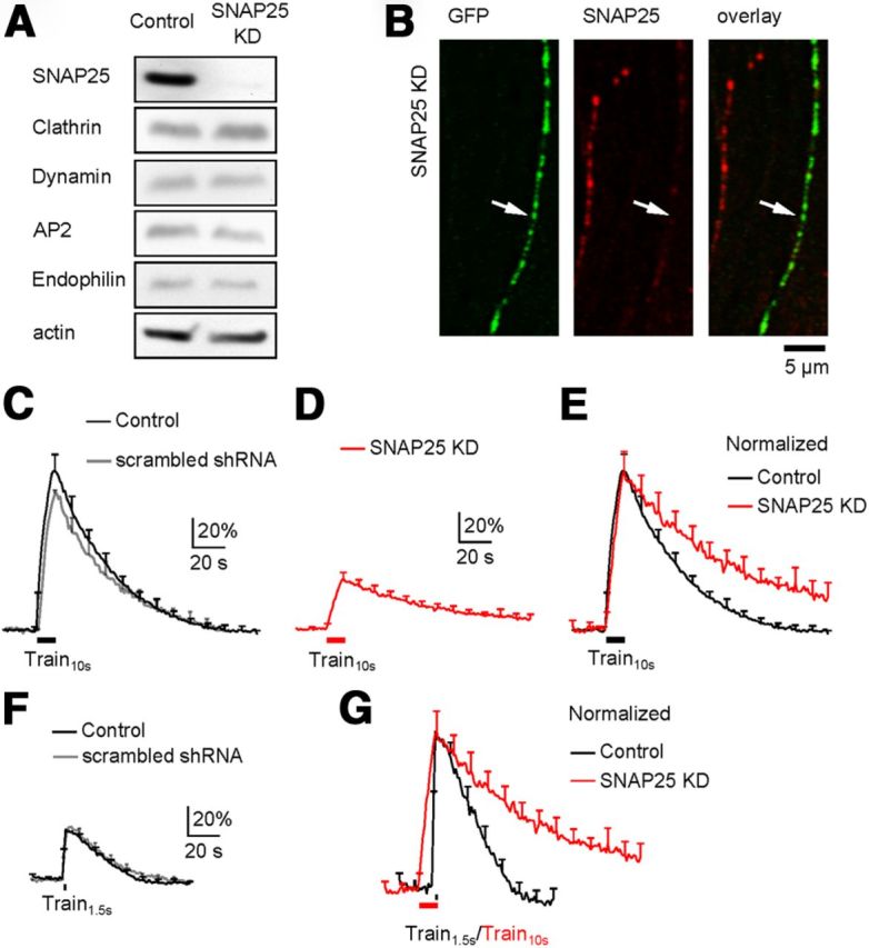
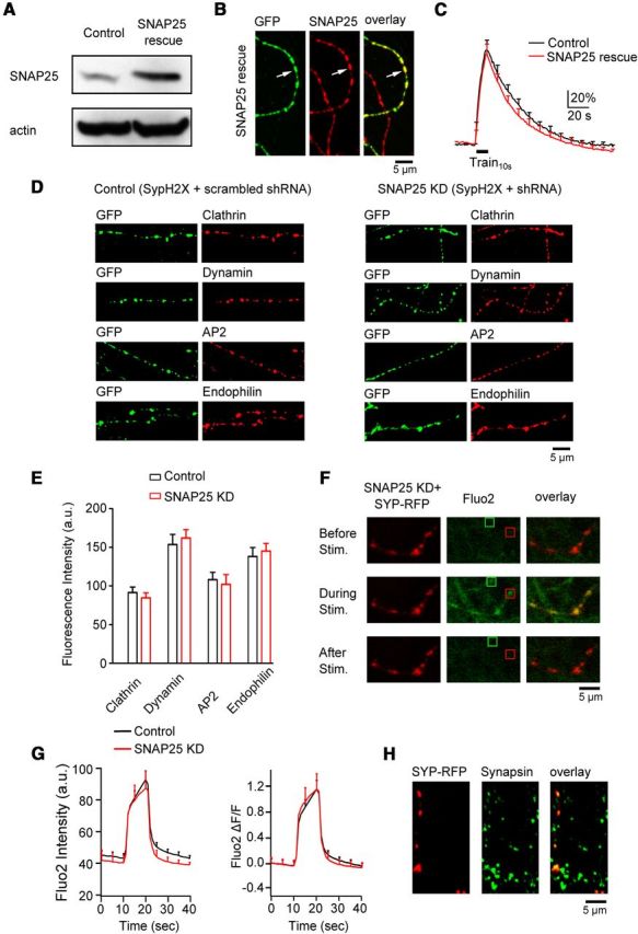
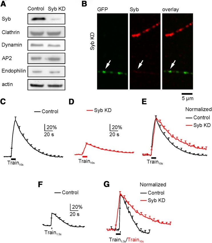
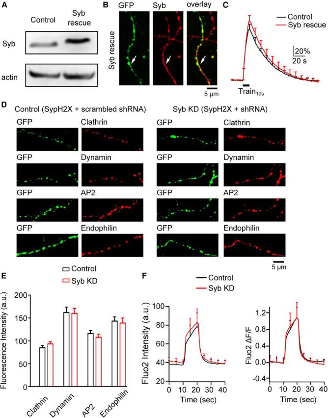
SNAP25, an essential component of the soluble NSF (N-ethylmaleimide-sensitive factor) attachment protein receptor (SNARE) complex that mediates exocytosis, is not considered to play a role in endocytosis, which couples to exocytosis by retrieving a similar amount of exocytosed vesicles. By knocking down SNAP25 and imaging slow endocytosis at a conventional synapse, the rat cultured hippocampal synapse, we found that SNAP25 is involved in slow, clathrin-dependent endocytosis. With similar techniques, we found that not only SNAP25, but also synaptobrevin is involved in slow endocytosis. These results provide the first evidence showing the dual role of SNAP25 and synaptobrevin in both exocytosis and slow endocytosis at conventional synapses. Such a dual role may contribute to mediate the coupling between exocytosis and clathrin-dependent endocytosis at conventional synapses, a mechanism critical for the maintenance of synaptic transmission and the normal structure of nerve terminals.


Figures

Figure 1.
Figure 1.
SNAP25 knockdown inhibits endocytosis. A, Western blot of SNAP25, clathrin, dynamin, AP2, endophilin, and actin from PC12 cells in control (left) and in cells transfected with SNAP25 shRNA (KD, right). B, Immunostaining of SypH2X (antibody against GFP, left, green) and SNAP25 (middle, red) at neuronal branches with (arrow, green staining) or without (no green staining) transfection of SypH2X and SNAP25 shRNA (right: left and middle panels superimposed). C, The SypH2X signal induced by Train10s at control boutons transfected with SypH2X (black, n = 6 experiments) or with SypH2X and scrambled shRNA (gray, n = 4). Data are expressed as the percentage change over the baseline intensity, and plotted as mean + SEM (every 10 s, applies to all similar plots). D, The SypH2X signal induced by Train10s at boutons transfected with SypH2X and SNAP25 shRNA (n = 12 experiments, SNAP25 KD). E, Traces in C (black only) and D (red) are normalized to the peak amplitude and superimposed. F, The SypH2X signal induced by Train1.5s at control boutons transfected with only SypH2X (black, n = 7 experiments) or with SypH2X and scrambled shRNA (gray, n = 7). G, Traces in F (black only) and D (red) are normalized to the same amplitude and superimposed.
Figure 2.
Figure 2.
SNAP25 knockdown is specific. A, Western blot of SNAP25 and actin from PC12 cells in control and in cells transfected with SNAP25 shRNA and shRNA-resistant SNAP25 (SNAP25 rescue). B, Immunostaining of SypH2X (antibody against GFP, left, green) and SNAP25 (middle, red) at neuronal branches with (arrow, green) or without (no green staining) triple-transfection of SypH2X, SNAP25 shRNA, and shNRA-resistant SNAP25 (SNAP25 rescue). Right, Left and middle panels superimposed. C, The SypH2X signal induced by Train10s at boutons transfected with only SypH2X (black, control, n = 6 experiments) or with SypH2X, SNAP25 shRNA, and shNRA-resistant SNAP25 (SNAP25 rescue, red, n = 8 experiments). D, Samples of immunostaining against GFP (green, recognizing SypH2X, first and third columns), clathrin, dynamin, AP2, and endophilin (red, second and fourth columns) at boutons transfected with scrambled shRNA and SypH2X (first and second columns, control) or with SNAP25 shRNA and SypH2X (third and fourth columns, SNAP25 KD). E, The immunostaining intensity of clathrin, dynamin, AP2, and endophilin at boutons transfected with scrambled shRNA and SypH2X (control, black) or with SNAP25 shRNA and SypH2X (SNAP25 KD, red). Each group was taken from 2 transfections (clathrin: control, n = 126 boutons; KD, n = 97 boutons; dynamin: control, n = 97 boutons; KD, n = 108 boutons; AP2: control, n = 84 boutons; KD, n = 86 boutons; endophilin: control, n = 91 boutons; KD, n = 95 boutons). Control and knockdown were not statistically significant for each protein. F, Sampled images showing fluo2 signal (calcium indicator, middle column, green) in neuronal branches with or without transfection of SNAP25 shRNA and synaptophysin-RFP (Syp-RFP, left column) at 10 s before (top), during (middle), and 20 s after (bottom) Train10s. Transfection was recognized as red (Syp-RFP) branches. Red and green squares show examples of stimulation-induced fluo2 puncta with and without transfection, respectively. Right, Left and middle columns superimposed. G, Fluo2 fluorescence raw data (left, a.u.) and fractional changes (right, ΔF/F, fluorescence change divided by the mean of baseline fluorescence) induced by Train10s in control boutons (n = 6 experiments, black) and in boutons transfected with SNAP25 shRNA and Syp-RFP (n = 6 experiments, red). H, Antibody staining against RFP (recognizing Syp-RFP, left) and synapsin (middle, bouton marker, green) in a culture transfected with SNAP25 shRNA and Syp-RFP. Right, Left and middle panels superimposed.
Figure 3.
Figure 3.
Synaptobrevin knockdown inhibits endocytosis. The arrangements in A–G are the same as those in Figure 1A–G, respectively, except that SNAP25 in all legends and panels is replaced with synaptobrevin (or Syb). C, The trace is the same as the black trace in Figure 1C. D, n = 10 experiments from boutons transfected with SypH2X and Syb shRNA (Syb KD). F, The trace is the same as the black trace in Figure 1F.
Figure 4.
Figure 4.
Synaptobrevin knockdown is specific. The arrangements in A–E and F are the same as Figure 2A–E and Figure 2G, respectively, except that SNAP25 in all legends and panels is replaced with synaptobrevin (or Syb). C, Control, n = 6 experiments; Syb rescue, n = 6 experiments). E, Each group was taken from 2 transfections (clathrin: control, n = 125 boutons; KD, n = 141 boutons; dynamin: control, n = 110 boutons; KD, n = 134 boutons; AP2: control, n = 106 boutons; KD, n = 95 boutons; endophilin: control, n = 120 boutons; KD, n = 128 boutons). F, control, n = 6 experiments; Syb KD, n = 6 experiments.

References

    1. Balaji J, Armbruster M, Ryan TA. Calcium control of endocytic capacity at a CNS synapse. J Neurosci. 2008;28:6742–6749. doi: 10.1523/JNEUROSCI.1082-08.2008. - DOI - PMC - PubMed
    1. Bronk P, Deák F, Wilson MC, Liu X, Südhof TC, Kavalali ET. Differential effects of SNAP-25 deletion on Ca2+-dependent and Ca2+-independent neurotransmission. J Neurophysiol. 2007;98:794–806. doi: 10.1152/jn.00226.2007. - DOI - PubMed
    1. Deák F, Schoch S, Liu X, Südhof TC, Kavalali ET. Synaptobrevin is essential for fast synaptic-vesicle endocytosis. Nat Cell Biol. 2004;6:1102–1108. doi: 10.1038/ncb1185. - DOI - PubMed
    1. Dittman J, Ryan TA. Molecular circuitry of endocytosis at nerve terminals. Ann Rev Cell Dev Biol. 2009;25:133–160. doi: 10.1146/annurev.cellbio.042308.113302. - DOI - PubMed
    1. Galas MC, Chasserot-Golaz S, Dirrig-Grosch S, Bader MF. Presence of dynamin–syntaxin complexes associated with secretory granules in adrenal chromaffin cells. J Neurochem. 2000;75:1511–1519. - PubMed

Publication types

MeSH terms

LinkOut - more resources