. 2013 Jul;162(3):1420-33.
doi: 10.1104/pp.113.215020. Epub 2013 May 22.

Functional implication of β-carotene hydroxylases in soybean nodulation

Affiliations

Functional implication of β-carotene hydroxylases in soybean nodulation

Yun-Kyoung Kim et al. Plant Physiol. 2013 Jul.

Abstract

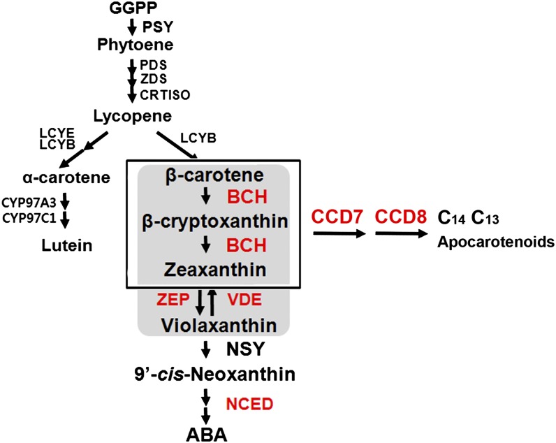
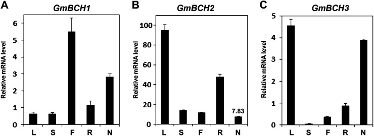
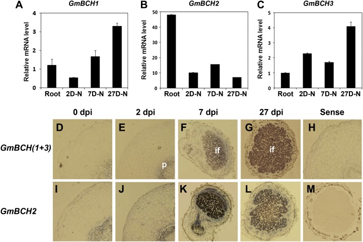
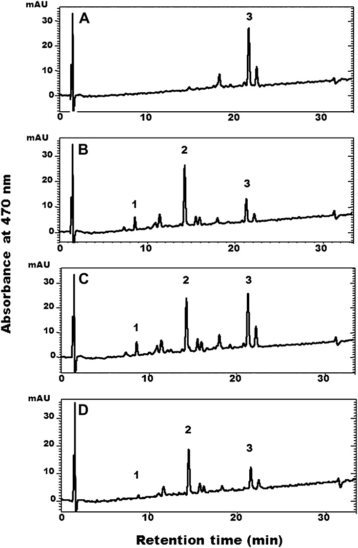
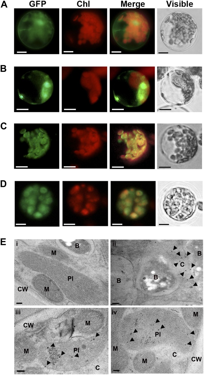
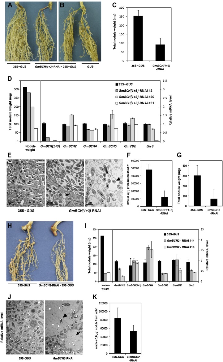
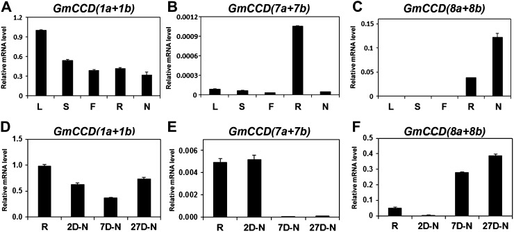
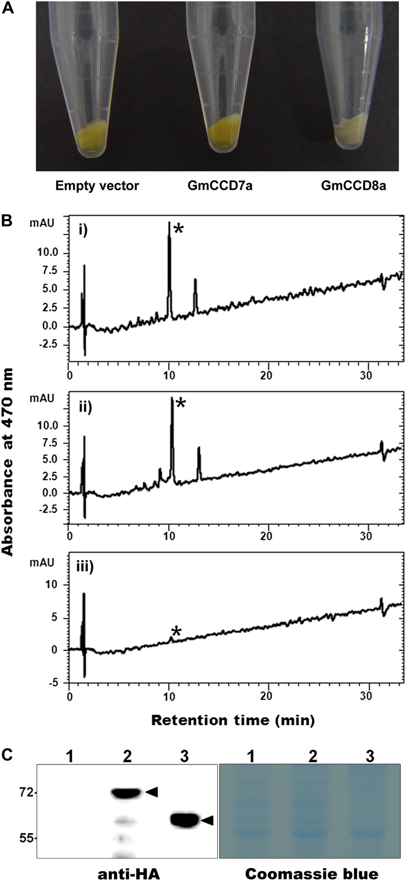
Legume-Rhizobium spp. symbiosis requires signaling between the symbiotic partners and differential expression of plant genes during nodule development. Previously, we cloned a gene encoding a putative β-carotene hydroxylase (GmBCH1) from soybean (Glycine max) whose expression increased during nodulation with Bradyrhizobium japonicum. In this work, we extended our study to three GmBCHs to examine their possible role(s) in nodule development, as they were additionally identified as nodule specific, along with the completion of the soybean genome. In situ hybridization revealed the expression of three GmBCHs (GmBCH1, GmBCH2, and GmBCH3) in the infected cells of root nodules, and their enzymatic activities were confirmed by functional assays in Escherichia coli. Localization of GmBCHs by transfecting Arabidopsis (Arabidopsis thaliana) protoplasts with green fluorescent protein fusions and by electron microscopic immunogold detection in soybean nodules indicated that GmBCH2 and GmBCH3 were present in plastids, while GmBCH1 appeared to be cytosolic. RNA interference of the GmBCHs severely impaired nitrogen fixation as well as nodule development. Surprisingly, we failed to detect zeaxanthin, a product of GmBCH, or any other carotenoids in nodules. Therefore, we examined the possibility that most of the carotenoids in nodules are converted or cleaved to other compounds. We detected the expression of some carotenoid cleavage dioxygenases (GmCCDs) in wild-type nodules and also a reduced amount of zeaxanthin in GmCCD8-expressing E. coli, suggesting cleavage of the carotenoid. In view of these findings, we propose that carotenoids such as zeaxanthin synthesized in root nodules are cleaved by GmCCDs, and we discuss the possible roles of the carotenoid cleavage products in nodulation.


Figures

Figure 1.
Figure 1.
The biosynthetic pathway of carotenoids in plants. GGPP, Geranylgeranyl diphosphate; PSY, phytoene synthase; PDS, phytoene desaturase; ZDS, ζ-carotene desaturase; CRTISO, carotene isomerase; LCYB, lycopene β-cyclase; CYP97A3 and CYP97C1, cytochrome P450 enzymes; NSY, neoxanthin synthase; LCYE, lycopene ε-cyclase; CRTR-E, ε-carotene hydroxylase. Enzymes in red were examined in this study.
Figure 2.
Figure 2.
Expression of the soybean BCHs GmBCH1, GmBCH2, and GmBCH3 in soybean tissues, including 27-d-old nodules. Transcript levels were determined by real-time RT-PCR and normalized with the geometric mean of three reference genes (GmELF1b, GmActin2/GmActin7, and Ubiquitin; Vandesompele et al., 2002). Data are representative of three independent experiments. Error bars represent sd (n = 3). L, Leaf; S, stem; F, flower; R, root; N, nodule.
Figure 3.
Figure 3.
Expression of GmBCHs during nodulation. A to C, Expression of GmBCH1 (A), GmBCH2 (B), and GmBCH3 (C) during nodule development. Transcript levels were determined by real-time RT-PCR and normalized. Expression levels are shown as means and sd of three independent experiments. 2D-N, Two-day-old nodule; 7D-N, 7-d-old nodule; 27D-N, 27-d-old nodule. D to M, In situ hybridization of GmBCHs during nodule development. Sections from 0 (roots), 2, 7, and 27 d post inoculation (dpi) were hybridized with antisense GmBCH(1+3) (D–G) and GmBCH2 (I–L) riboprobes. Sections from 0 (roots) and 27 dpi were hybridized with sense GmBCH(1+3) (H) and GmBCH2 (M) riboprobes as negative controls. if, Infected region; p, Pericycle.
Figure 4.
Figure 4.
HPLC analysis of carotenoids extracted from β-carotene-producing E. coli transformed with GmBCH1, GmBCH2, or GmBCH3. pACCAR16ΔcrtX-containing E. coli harboring pUC19 alone as a negative control (A) or harboring pGmBCH1 (B), pGmBCH2 (C), or pGmBCH3 (D) was used for carotenoid analysis. Peak 1, Zeaxanthin; peak 2, β-cryptoxanthin; peak 3, β-carotene. mAU, Milliabsorbance units.
Figure 5.
Figure 5.
Subcellular localization of GmBCHs in Arabidopsis protoplasts and soybean nodules. A to D, Arabidopsis protoplasts were transfected with control vector (35S-GFP; A), GmBCH1-GFP (B), GmBCH2-GFP (C), or GmBCH3-GFP (D). Bars = 10 μm. E, GmBCHs were localized by EM immunogold labeling with an anti-GFP antiserum in soybean nodules transformed with empty vector (pCAMBIA3301; i), GmBCH1-GFP (ii), GmBCH2-GFP (iii), or GmBCH3-GFP (iv). Gold particles (10 nm) are indicated by arrowheads. B, Bacteroids; C, cytoplasm; CW, cell wall; M, mitochondria; Pl, plastid. Bars = 100 nm.
Figure 6.
Figure 6.
Nodule development on transgenic hairy roots expressing an RNAi construct against both GmBCH1 and GmBCH3. A, Nodules formed on transgenic hairy roots containing pCAMBIA1304 alone (control; 35S-GUS; left plant) or the GmBCH(1+3)-RNAi cloned in pCAMBIA1304 (right plant). After GUS assay, only GUS-positive (transgenic) hairy roots were inoculated with B. japonicum (K599). These experiments were repeated three times, and representative results are shown. In each experiment, five to seven plants were used per construct. B, A GUS-negative (untransformed control) hairy root is shown on the right. C, Total nodule weights (mg) of control (35S-GUS) and GmBCH(1+3)-RNAi plants were measured and are shown as means and sd of three independent experiments. D, Transcript levels are shown in nodules from a transgenic control plant (35S-GUS) and three representative transgenic plants: GmBCH(1+3)-RNAi 2, GmBCH(1+3)-RNAi 20, and GmBCH(1+3)-RNAi 21. RNAi plant 2 had almost the same nodule weight as the control, RNAi plant 20 had a reduced nodule weight, and RNAi plant 21 had a drastically reduced nodule weight. Transcript levels of GmBCH(1+3), GmBCH2, GmBCH4, GmBCH5, GmVDE, and Lbc3 were determined by real-time RT-PCR in controls (35S-GUS) and three GmBCH(1+3)-RNAi nodules and normalized. Expression levels are shown as means and sd of three independent experiments. E, EM images of 27-d-old control (35S-GUS) and GmBCH(1+3)-RNAi plants. GmBCH(1+3)-RNAi nodules often contained empty vesicles (arrow) and bacteroids outside the symbiosomes (arrowhead). F, Nitrogenase activities were measured by the acetylene reduction assay, and data are averaged from three independent experiments. G, Total nodule weights (mg) of control (35S-GUS) and GmBCH2-RNAi plants were measured and are shown as means and sd of three independent experiments. H, Nodules formed on transgenic hairy roots containing pCAMBIA1304 alone (control; 35S-GUS; left plant) or the GmBCH2-RNAi cloned in pCAMBIA1304 (right plant). After GUS assay, nodules were formed as in A. I, Transcript levels in nodules from one transgenic control plant (35S-GUS) and two differentially repressed transgenic plants (GmBCH2-RNAi 14 and GmBCH2-RNAi 16) are shown. Transcript levels of GmBCH2, GmBCH(1+3), GmBCH4, GmBCH5, GmVDE, and Lbc3 were determined by real-time RT-PCR and normalized. Expression levels are shown as means and sd of three independent experiments. J, EM images of 27-d-old control (35S-GUS) and GmBCH2-RNAi nodules. GmBCH2-RNAi nodules often contained empty vesicles (arrow) and bacteroids outside the symbiosomes (arrowhead). K, Nitrogenase activities were measured by the acetylene reduction assay as in F.
Figure 7.
Figure 7.
Expression of GmZEPs, GmVDEs, and GmNCED1s in soybean. A to C, Expression of GmZEPs, GmVDEs, and GmNCED1s in different tissues. RNA was extracted from different tissues, including 27-d-old nodules of soybean. D to F, Expression of GmZEPs, GmVDEs, and GmNCED1s during nodule development. RNAs were extracted from roots and 2-, 7-, and 27-d-old nodules, and transcript levels were determined by real-time RT-PCR and normalized. Expression of GmZEP1 and GmZEP2 was examined simultaneously using primers for DNA regions of high identity, while the expression of GmZEP3, a gene with low overall homology to other GmZEPs, was measured separately and is shown in Supplemental Figure S10. Expression of GmVDE1 and GmVDE2 was also examined simultaneously using primers for the DNA regions of high identity. Expression of GmNCED1a and GmNCED1b was also examined simultaneously using primers for the DNA regions of high identity. Data are from three independent experiments. L, Leaf; S, stem; F, flower; R, root; N, nodule; 2D-N, 2-d-old nodule; 7D-N, 7-d-old nodule; 27D-N, 27-d-old nodule.
Figure 8.
Figure 8.
Expression of GmCCDs in soybean. A to C, Expression of GmCCD1s, GmCCD7s, and GmCCD8s in different tissues. RNA was extracted from various tissues, including 27-d-old nodules of soybean. D to F, Expression of GmCCD1s, GmCCD7s, and GmCCD8s during nodulation. RNAs were extracted from roots and 2-, 7-, and 27-d-old nodules, and transcript levels were determined by real-time RT-PCR and normalized. Expression levels are shown as means of three independent experiments. L, Leaf; S, stem; F, flower; R, root; N, nodule; 2D-N, 2-d-old nodule; 7D-N, 7-d-old nodule; 27D-N, 27-d-old nodule.
Figure 9.
Figure 9.
Functional assays of GmCCD7 and GmCCD8 in E. coli. A, Expression of GmCCD7a (middle) or GmCCD8a (right) in E. coli strains that carry pAC-zeaxanthin and accumulate zeaxanthin. A zeaxanthin-accumulating E. coli strain with empty vector alone (left) served as a negative control. B, HPLC analysis of carotenoids extracted from zeaxanthin-accumulating E. coli cells expressing GmCCD7a (ii) or GmCCD8a (iii) or with empty vector (i). The zeaxanthin peak is indicated by the asterisks. mAU, Milliabsorbance units. C, Proteins from zeaxanthin-accumulating E. coli cells expressing HA-GmCCD7 (lane 2) and HA-GmCCD8 (lane 3) or with empty vector (lane 1) were isolated (right panel) and immunoblotted with hemagglutinin (HA) antibody (left panel). Proteins of the expected sizes corresponding to GmCCD7 (70 kD) and GmCCD8 (60 kD) were detected. Immunodetected bands are indicated by arrowheads. [See online article for color version of this figure.]

References

    1. Alder A, Holdermann I, Beyer P, Al-Babili S. (2008) Carotenoid oxygenases involved in plant branching catalyse a highly specific conserved apocarotenoid cleavage reaction. Biochem J 416: 289–296 - PubMed
    1. Auldridge ME, Block A, Vogel JT, Dabney-Smith C, Mila I, Bouzayen M, Magallanes-Lundback M, DellaPenna D, McCarty DR, Klee HJ. (2006a) Characterization of three members of the Arabidopsis carotenoid cleavage dioxygenase family demonstrates the divergent roles of this multifunctional enzyme family. Plant J 45: 982–993 - PubMed
    1. Auldridge ME, McCarty DR, Klee HJ. (2006b) Plant carotenoid cleavage oxygenases and their apocarotenoid products. Curr Opin Plant Biol 9: 315–321 - PubMed
    1. Booker J, Auldridge M, Wills S, McCarty D, Klee H, Leyser O. (2004) MAX3/CCD7 is a carotenoid cleavage dioxygenase required for the synthesis of a novel plant signaling molecule. Curr Biol 14: 1232–1238 - PubMed
    1. Bouvier F, Isner JC, Dogbo O, Camara B. (2005) Oxidative tailoring of carotenoids: a prospect towards novel functions in plants. Trends Plant Sci 10: 187–194 - PubMed

Publication types

MeSH terms

LinkOut - more resources