

Detection of JC virus-specific immune responses in a novel humanized mouse model
- PMID: 23700470
- PMCID: PMC3658964
- DOI: 10.1371/journal.pone.0064313
Detection of JC virus-specific immune responses in a novel humanized mouse model
Abstract
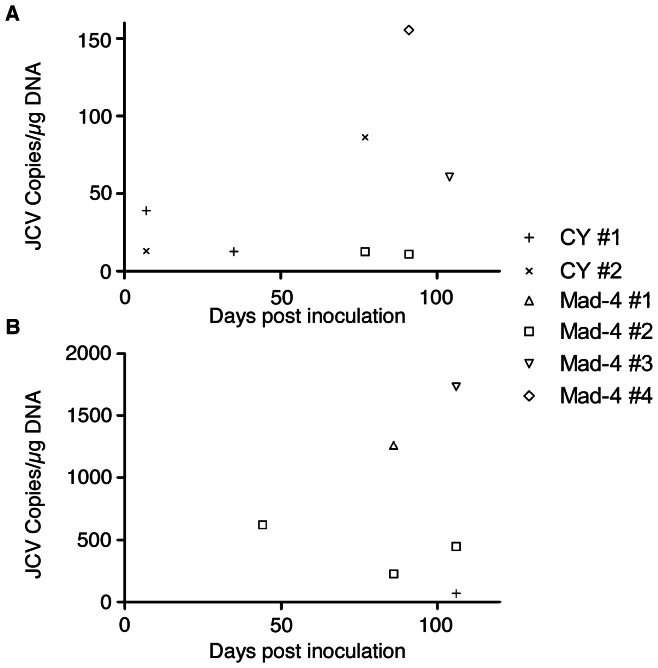
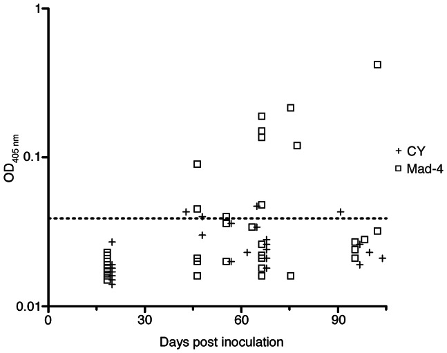
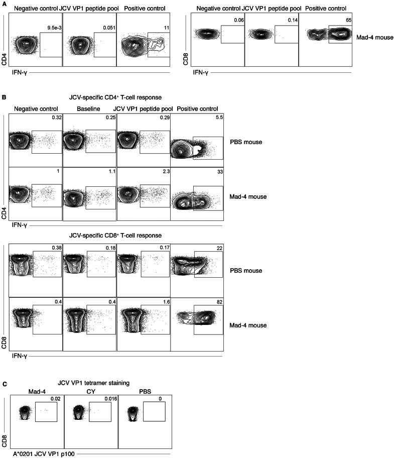
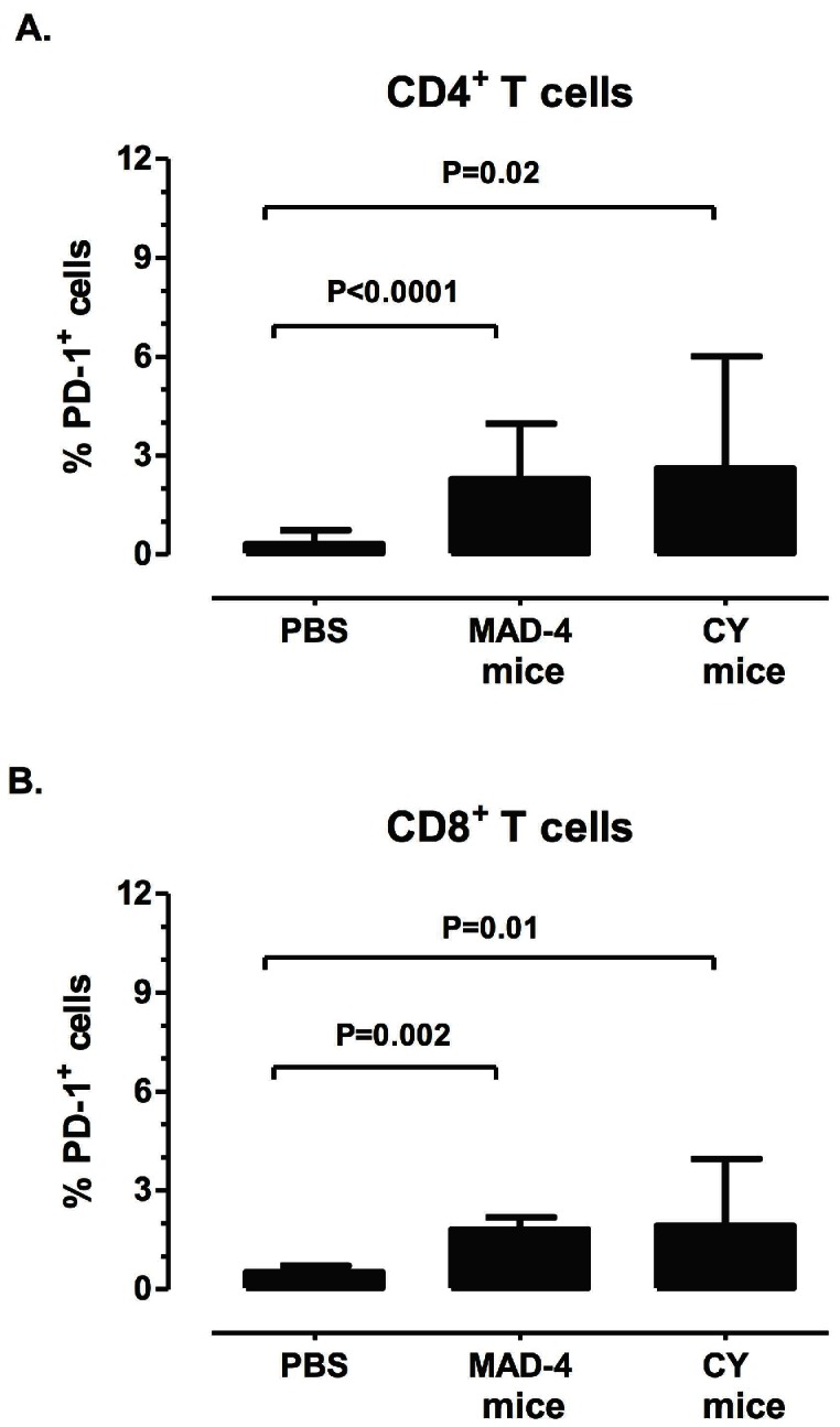
Progressive Multifocal Leukoencephalopathy (PML) is an often fatal disease caused by the reactivation of the JC virus (JCV). Better understanding of viral-host interactions has been hampered by the lack of an animal model. Engrafting NOD/SCID/IL-2-Rg (null) mice with human lymphocytes and thymus, we generated a novel animal model for JCV infection. Mice were inoculated with either a PML isolate, JCV Mad-4, or with JCV CY, found in the kidney and urine of healthy individuals. While mice remained asymptomatic following inoculation, JCV DNA was occasionally detected in both the blood and the urine compartments. Mice generated both humoral and cellular immune responses against JCV. Expressions of immune exhaustion marker, PD-1, on lymphocytes were consistent with response to infection. Using this model we present the first in vivo demonstration of virological and immunological differences between JCV Mad-4 and CY. This model may prove valuable for studying JCV host immune responses.
Conflict of interest statement
Figures
References
-
- Egli A, Infanti L, Dumoulin A, Buser A, Samaridis J, et al. (2009) Prevalence of Polyomavirus BK and JC Infection and Replication in 400 Healthy Blood Donors. J Infect Dis 199 6: 837–846. - PubMed
-
- Markowitz RB, Thompson HC, Mueller JF, Cohen JA, Dynan WS (1993) Incidence of BK virus and JC virus viruria in human immunodeficiency virus-infected and -uninfected subjects. J Infect Dis 167: 13–20. - PubMed
-
- Du Pasquier RA, Kuroda MJ, Zheng Y, Jean-Jacques J, Letvin NL, et al. (2004) A prospective study demonstrates an association between JC virus-specific cytotoxic T lymphocytes and the early control of progressive multifocal leukoencephalopathy. Brain 127: 1970–1978. - PubMed
Publication types
MeSH terms
Substances
Grants and funding
- T32 AI007061/AI/NIAID NIH HHS/United States
- K24 NS 060950/NS/NINDS NIH HHS/United States
- NS R56 041198/NS/NINDS NIH HHS/United States
- P30 AI60354/AI/NIAID NIH HHS/United States
- R01 NS074995/NS/NINDS NIH HHS/United States
- K08 NS064215/NS/NINDS NIH HHS/United States
- R01 NS 047029/NS/NINDS NIH HHS/United States
- NS 074995/NS/NINDS NIH HHS/United States
- K08 NS 064215-01/NS/NINDS NIH HHS/United States
- P30 AI060354/AI/NIAID NIH HHS/United States
- R01 NS047029/NS/NINDS NIH HHS/United States
- K24 NS060950/NS/NINDS NIH HHS/United States
LinkOut - more resources
Full Text Sources
Other Literature Sources
