A novel biological approach to treat chondromalacia patellae
- PMID: 23700485
- PMCID: PMC3659098
- DOI: 10.1371/journal.pone.0064569
A novel biological approach to treat chondromalacia patellae
Abstract
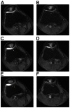
Mesenchymal stem cells from several sources (bone marrow, synovial tissue, cord blood, and adipose tissue) can differentiate into variable parts (bones, cartilage, muscle, and adipose tissue), representing a promising new therapy in regenerative medicine. In animal models, mesenchymal stem cells have been used successfully to regenerate cartilage and bones. However, there have been no follow-up studies on humans treated with adipose-tissue-derived stem cells (ADSCs) for the chondromalacia patellae. To obtain ADSCs, lipoaspirates were obtained from lower abdominal subcutaneous adipose tissue. The stromal vascular fraction was separated from the lipoaspirates by centrifugation after treatment with collagenase. The stem-cell-containing stromal vascular fraction was mixed with calcium chloride-activated platelet rich plasma and hyaluronic acid, and this ADSCs mixture was then injected under ultrasonic guidance into the retro-patellar joints of all three patients. Patients were subjected to pre- and post-treatment magnetic resonance imaging (MRI) scans. Pre- and post-treatment subjective pain scores and physical therapy assessments measured clinical changes. One month after the injection of autologous ADSCs, each patient's pain improved 50-70%. Three months after the treatment, the patients' pain improved 80-90%. The pain improvement persisted over 1 year, confirmed by telephone follow ups. Also, all three patients did not report any serious side effects. The repeated magnetic resonance imaging scans at three months showed improvement of the damaged tissues (softened cartilages) on the patellae-femoral joints. In patients with chondromalacia patellae who have continuous anterior knee pain, percutaneous injection of autologous ADSCs may play an important role in the restoration of the damaged tissues (softened cartilages). Thus, ADSCs treatment presents a glimpse of a new promising, effective, safe, and non-surgical method of treatment for chondromalacia patellae.
Conflict of interest statement
Figures


References
-
- Brody LT, Thein JM (1998) Nonoperative treatment for patellofemoral pain. J Orthop Sports Phys Ther 28: 336–344. - PubMed
-
- Wittstein JR, O'Brien SD, Vinson EN, Garrett WE Jr (2009) MRI evaluation of anterior knee pain: predicting response to nonoperative treatment. Skeletal Radiol 38: 895–901. - PubMed
-
- Centeno CJ, Busse D, Kisiday J, Keohan C, Freeman M, et al. (2008) Increased knee cartilage volume in degenerative joint disease using percutaneously implanted, autologous mesenchymal stem cells. Pain Physician 11: 343–353. - PubMed
-
- Pak J (2012) Autologous adipose tissue-derived stem cells induce persistent bone-like tissue in osteonecrotic femoral heads. Pain Physician 15: 75–85. - PubMed
-
- Korean Food and Drug Administration (KFDA) (2009) Cell therapy: Rules and Regulations, Chapter 2, Section 14. Seoul (Korea): KFDA. 4 p.
Publication types
MeSH terms
LinkOut - more resources
Full Text Sources
Other Literature Sources

