. 2012 Jan 1;1(1):13-24.
doi: 10.4161/adip.18951.

Brown adipose tissue: Recent insights into development, metabolic function and therapeutic potential

Affiliations

Brown adipose tissue: Recent insights into development, metabolic function and therapeutic potential

Kristy Townsend et al. Adipocyte. .

Abstract

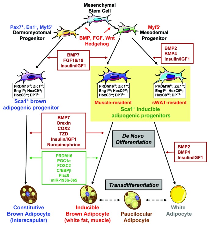
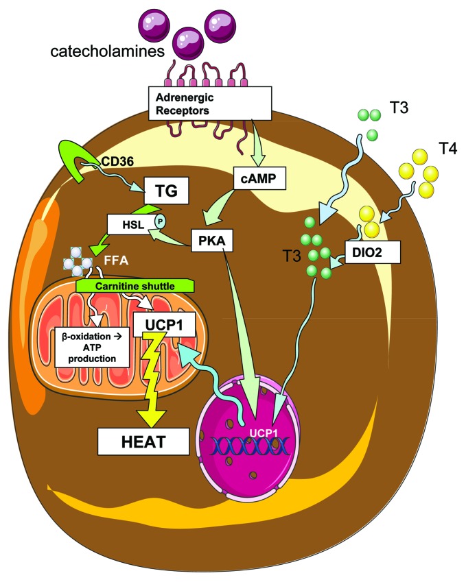
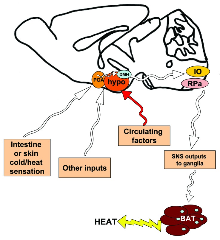
Obesity is currently a global pandemic, and is associated with increased mortality and co-morbidities including many metabolic diseases. Obesity is characterized by an increase in adipose mass due to increased energy intake, decreased energy expenditure, or both. While white adipose tissue is specialized for energy storage, brown adipose tissue has a high concentration of mitochondria and uniquely expresses uncoupling protein 1, enabling it to be specialized for energy expenditure and thermogenesis. Although brown fat was once considered only necessary in babies, recent morphological and imaging studies have provided evidence that, contrary to prior belief, this tissue is present and active in adult humans. In recent years, the topic of brown adipose tissue has been reinvigorated with many new studies regarding brown adipose tissue differentiation, function and therapeutic promise. This review summarizes the recent advances, discusses the emerging questions and offers perspective on the potential therapeutic applications targeting this tissue.

Keywords: BATokine; UCP1; adipogenesis; brown adipose tissue; browning; mitochondria; thermogenesis.


Figures

None
Figure 1. Regulation of brown adipocyte development. Brown adipocytes located in different anatomical locations of the body arise from different developmental origins. While the Pax7+/En1+/Myf5+ dermomyotome progenitor gives rise to interscapular brown fat, a distinct myf5 tissue resident progenitor serves as the common precursor for white adipocytes and systemic brown adipocytes. With the stimulation of appropriate developmental cues, these progenitors become committed to the adipocyte lineage. The Sca-1+ progenitor cells isolated from interscapular brown fat serve as constitutively committed brown fat precursors, and Sca-1+ progenitor cells from skeletal muscle and subcutaneous white fat are highly inducible to become mature brown adipocytes. These precursors possess unique molecular signatures that allow designation of the distinction of cellular origin. Agents that can promote brown adipocyte differentiation include norepinephrine, insulin/IGF1, thiazolidinedione (TZD), cyclooxygenase 2 (COX2), orexin, BMP7 and others. At the molecular level, a number of transcriptional/post-transcriptional regulators have been shown to specify or enhance brown fat phenotype, such as PRDM16, PGC1α, FOXC2, C/EBPβ, Plac8 and miR-193b-365. The brown adipocytes in white fat may come from de novo differentiation and/or transdifferentiation. Dashed lines in this figure indicate links that are only partially established.
None
Figure 2. Brown adipose activity in response to sympathetic input and thyroid hormone. Catecholamines bind to adrenergic receptors on the surface of brown adipocytes, initiating signaling cascades that include cAMP and protein kinase A (PKA), which then phosphorylates and activates the enzyme hormone sensitive lipase (HSL), which then cleaves triglycerides (TG) into free fatty acids (FFA). Triglycerides enter the cell by uptake of triglyceride rich lipoproteins via CD36 transport, and fatty acids then enter the mitochondria through the carnitine shuttle. Mitochondrial fatty acids may be oxidized via β-oxidation, or serve to activate UCP1 thermogenesis. Additionally, thyroid hormones T3 and T4 enter the cell, and T4 is further converted to T3 by type 2 deiodinase (DIO2). T3 is then able to affect mitochondrial activity and nuclear transcription of genes that affect energenesis, including UCP1. PKA also affects nuclear transcription of UCP1, a protein which acts in the mitochondria to uncouple oxidative phosphorylation from ATP production, resulting in heat generation.
None
Figure 3. Simplified schematic of neural pathways in the mouse brain which affect sympathetic outflow to brown adipose tissue. Using rodent models, neuroscientists have begun to identify which neural pathways are involved in signaling temperature status (such as cold) to the brain, followed by sympathetic stimulation of brown adipose tissue in order to initiate thermogenesis. Some of these findings are summarized in this mid-sagittal view of a mouse brain, but for a complete review see Morrison et al. Cold temperature is sensed by the pre-optic area (POA), rostral to the hypothalamus (hypo). The POA sends signals to the hypothalamus, including the dorsomedial hypothalamus (DMH). Other hypothalamic nuclei are also involved in relaying various signals related to energy status, in response to various neural inputs and circulating factors. Neural outputs from the hypothalamus reach the inferior olive and GABAergic centers in the raphe pallidus (RPa) in the medulla of the brain stem. From here, sympathetic outputs are activated and send afferents to the sympathetic ganglia, followed by the brown adipose tissue, where catecholamine neurotransmitters are released from sympathetic nerve terminals, to act on adrenergic receptors there. White arrows represent neural pathways.

References

    1. Caballero B. The global epidemic of obesity: an overview. Epidemiol Rev. 2007;29:1–5. doi: 10.1093/epirev/mxm012. - DOI - PubMed
    1. Gesta S, Tseng YH, Kahn CR. Developmental origin of fat: tracking obesity to its source. Cell. 2007;131:242–56. doi: 10.1016/j.cell.2007.10.004. - DOI - PubMed
    1. Cannon B, Nedergaard J. Metabolic consequences of the presence or absence of the thermogenic capacity of brown adipose tissue in mice (and probably in humans) Int J Obes (Lond) 2010;34(Suppl 1):S7–16. doi: 10.1038/ijo.2010.177. - DOI - PubMed
    1. Nedergaard J, Cannon B. The changed metabolic world with human brown adipose tissue: therapeutic visions. Cell Metab. 2010;11:268–72. doi: 10.1016/j.cmet.2010.03.007. - DOI - PubMed
    1. Nedergaard J, Bengtsson T, Cannon B. Three years with adult human brown adipose tissue. Ann N Y Acad Sci. 2010;1212:E20–36. doi: 10.1111/j.1749-6632.2010.05905.x. - DOI - PubMed