Review
. 2014 Feb;10(1):38-56.
doi: 10.2174/1573403x113099990005.

Pathophysiology of coronary collaterals

Affiliations
Review

Pathophysiology of coronary collaterals

Michael Stoller et al. Curr Cardiol Rev. 2014 Feb.

Abstract

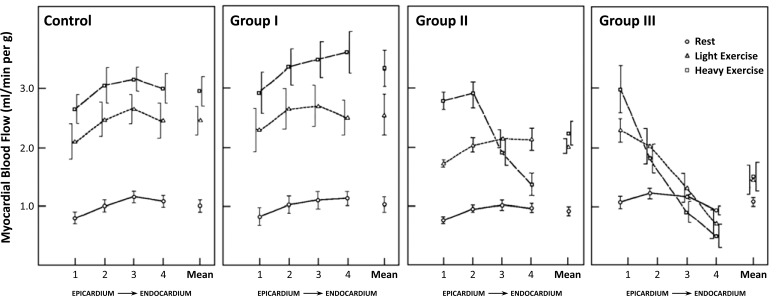
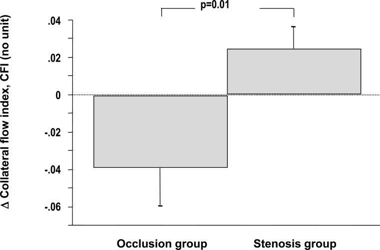
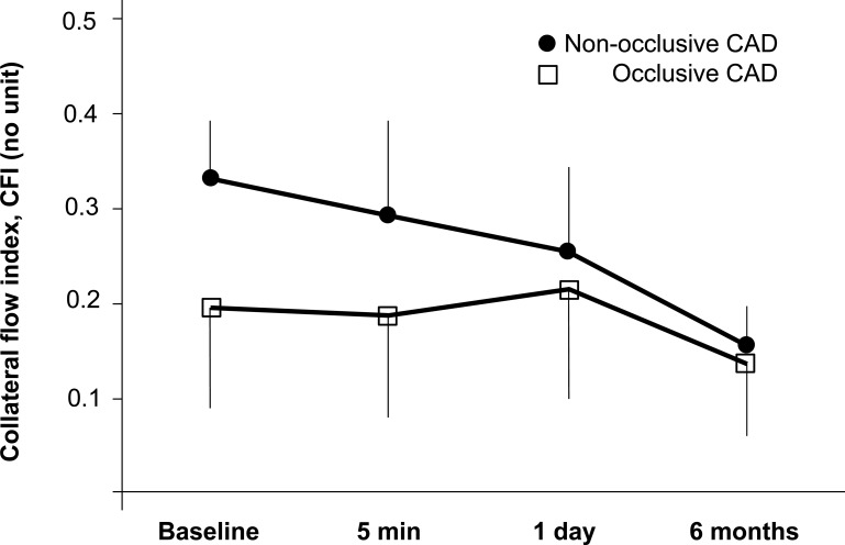
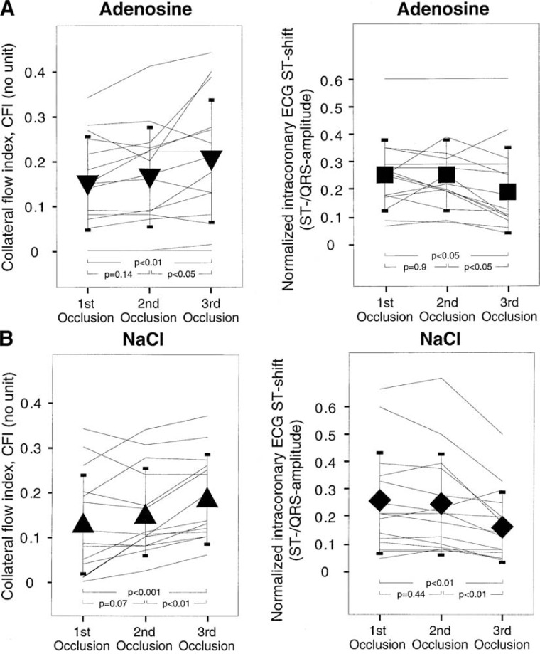
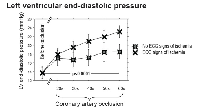
While the existence of structural adaptation of coronary anastomoses is undisputed, the potential of coronary collaterals to be capable of functional adaptation has been questioned. For many years, collateral vessels were thought to be rigid tubes allowing only limited blood flow governed by the pressure gradient across them. This concept was consistent with the notion that although collaterals could provide adequate blood flow to maintain resting levels, they would be unable to increase blood flow sufficiently in situations of increased myocardial oxygen demand. However, more recent studies have demonstrated the capability of the collateral circulation to deliver sufficient blood flow even during exertion or pharmacologic stress. Moreover, it has been shown that increases in collateral flow could be attributed directly to collateral vasomotion. This review summarizes the pathophysiology of the coronary collateral circulation, ie the functional adapation of coronary collaterals to acute alterations in the coronary circulation.


Figures

Fig. (1)
Fig. (1)
Myocardial blood flow in the collateral-dependent myocardial region of 14 dogs after 1 month after placement of an ameroid constrictor of the left circumflex artery (group I to III) and in 7 normal dogs (control). Data (means ± SE) are shown for 4 transmural layers from epicardium to endocardium at rest and during two levels of treadmill exercise. Group I shows collateralization sufficient to allow completely normal increase in myocardial blood flow during both levels of exercise. Group II shows subnormal increase in blood flow during light exercise and relative underperfusion during heavy exercise, expressing as transmural redistribution of flow towards the subepicardium. Group III shows collateralization sufficient only at rest, with already light exercise leading to redistribution towards the epicardium and heavy exercise leading to an actual decrease of myocardial blood levels in the inner layers to below resting levels (transmural steal). Dot inside symbol denotes significant differences from corresponding control (p<0.05). [Data from Bache and Schwartz [15]].
Fig. (2)
Fig. (2)
Individual changes (thin lines) of collateral flow index (collateral flow index; vertical axes) from the resting condition to the peak supine bicycle exercise condition in the group ‘rest first’ (left panel; triangular symbols) and vice versa in the group ‘exercise first’ (right panel; cross symbols). CFI increased during exercise in 24 patients and decreased in six patients (coronary steal). Thick lines indicate the mean CFI change between the resting and exercise condition. Error bars denote standard deviation. See text for further explanation. [Data from Togni et al. [25]].
Fig. (3)
Fig. (3)
Upper panel: Individual, collateral flow index values at baseline and during intravenous adenosine infusion for patients with poor and good collaterals. The triangles indicate mean values (± standard deviation). Bottom panel: Individual, absolute CFIp changes in response to adenosine (i.e. CFIp during hyperemia − CFIp at rest) for the groups with poor and good collaterals, respectively. CFI remained practically unchanged during adenosine infusion in the group with poor collaterals. In the group with good collaterals, mean CFI increased significantly during adenosine infusion, but the response was highly variable. CFIp = pressure derived collateral flow index. See text for further explanation [Data from Seiler et al. [38]].
Fig. (4)
Fig. (4)
Diagram depicting coronary collateral steal. At rest, microcirculation of myocardial region distal to stenotic conductance vessel maintains normal perfusion by compensatory predilatation. In situation illustrated, half of perfusion (50 mL/min [mL/′]) is provided by antegrade flow through stenotic vessel and half by collaterals (50 mL/min) from contralateral nonstenotic vessel. During hyperemia, capacity for further arteriolar dilatation in poststenotic region (left) is exhausted, whereas it is intact on contralateral collateral-supplying side. Thus, microvascular resistance is more reduced on collateral-providing side than on poststenotic side, thereby reducing flow via collaterals (to 25 mL/min in this example) and causing net myocardial perfusion during hyperemia to be less (ie, 85mL/min) than at resting conditions (ie, 100mL/min). [Data from Seiler et al. [56]].
Fig. (5)
Fig. (5)
Changes in collateral flow between the first and the second balloon occlusion after revascularization in patients with (occlusion group) and without a chronic total occlusion (stenosis group). While patients with a prior total coronary occlusion show a trend for collateral de-recruitment, patients with a prior stenosis show intact collateral recruitment. See text for further explanation. [Data from Pohl et al. [77]].
Fig. (6)
Fig. (6)
Behaviour of collateral blood flow before and after percutaneous coronary intervention for non-occlusive stenosis and chronic total occlusions. Patients with CTOs had a higher baseline CFI and a greater decline in CFI over time than those with non-occlusive lesions (p<0.0001) (error bars=1 SD). See text for further explanation. [Data from Perera et al. [79]].
Fig. (7)
Fig. (7)
Forest plot of risk ratios (RR) for restenosis after percutaneous coronary intervention. Restenosis is defined as ≥ 50% diameter stenosis. Patients with good collateralization show a significantly increased risk for restenosis compared to patients with poor coronary collateralization. Markers represent point estimates of risk ratios, marker size represents study weight in random effects meta-analysis. Horizontal bars indicate 95% confidence intervals. [Data from Meier et al. [94]].
Fig. (8)
Fig. (8)
Intraindividual changes in collateral flow (left panels) and in normalized ECG ST shift in the intracoronary ECG (right panels) during three consecutive balloon occlusion of each 2 minutes duration. Upper panels show data for the group pretreated with adenosine infusion, lower panels shows control group without adenosine pretreatment (NaCl infusion). Collateral flow shows a trend for an increase from the first to the second occlusion and a significant increase in the third balloon occlusion. ST shift in the intracoronary ECG is significantly lower only in the third balloon occlusion. See text for further explanation. [Data from Billinger et al. [113]].
Fig. (9)
Fig. (9)
Intraindividual LVEDP changes during coronary balloon occlusion in patients with sufficient (blue symbols) and insufficient collaterals (black symbols). LVEDP raises similarly at the start of the coronary occlusion in both groups, but reaches a plateau in patients with sufficient collaterals, whereas LVEDP in patients with insufficient collaterals continues to increase. See text for further explanation. [Data from de Marchi et al. [135]].

Similar articles

Cited by

References

    1. Feigl EO. Coronary physiology. Physiol Rev. 1983;63:1–205. - PubMed
    1. Gregg DE. Coronary Circulation in Health and Disease. Philadelphia PA. Lea & Febiger. 1950
    1. Hoffman JI, Spaan JA. Pressure-flow relations in coronary circulation. Physiol Rev. 1990;70:331–90. - PubMed
    1. Klocke FJ. Coronary blood flow in man. Prog Cardiovasc Dis. 1976;19:117–66. - PubMed
    1. Klocke FJ, Ellis AK. Control of coronary blood flow. Annu Rev Med. 1980;31:489–508. - PubMed

MeSH terms