

Mapping of conserved and species-specific antibody epitopes on the Ebola virus nucleoprotein
- PMID: 23702199
- PMCID: PMC3787873
- DOI: 10.1016/j.virusres.2013.05.004
Mapping of conserved and species-specific antibody epitopes on the Ebola virus nucleoprotein
Abstract
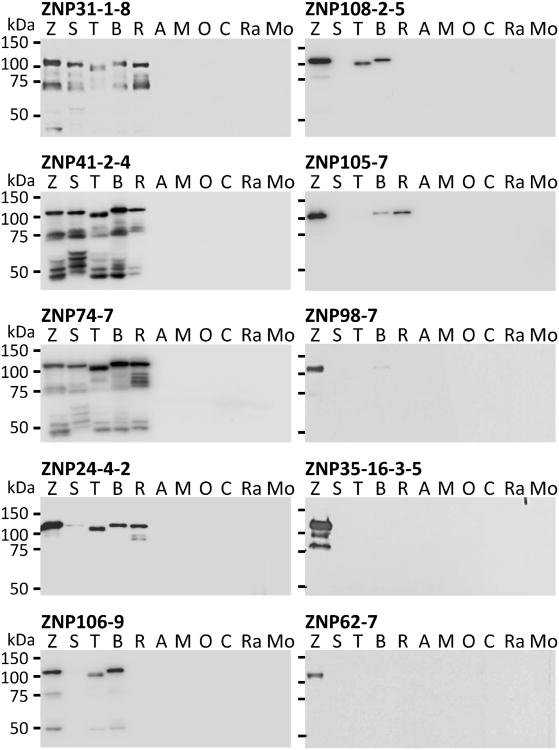
Filoviruses (viruses in the genus Ebolavirus and Marburgvirus in the family Filoviridae) cause severe haemorrhagic fever in humans and nonhuman primates. Rapid, highly sensitive, and reliable filovirus-specific assays are required for diagnostics and outbreak control. Characterisation of antigenic sites in viral proteins can aid in the development of viral antigen detection assays such immunochromatography-based rapid diagnosis. We generated a panel of mouse monoclonal antibodies (mAbs) to the nucleoprotein (NP) of Ebola virus belonging to the species Zaire ebolavirus. The mAbs were divided into seven groups based on the profiles of their specificity and cross-reactivity to other species in the Ebolavirus genus. Using synthetic peptides corresponding to the Ebola virus NP sequence, the mAb binding sites were mapped to seven antigenic regions in the C-terminal half of the NP, including two highly conserved regions among all five Ebolavirus species currently known. Furthermore, we successfully produced species-specific rabbit antisera to synthetic peptides predicted to represent unique filovirus B-cell epitopes. Our data provide useful information for the development of Ebola virus antigen detection assays.
Keywords: Antibody epitope; BDBV; Bundibugyo virus; EBOV; Ebola virus; GP; MARV; Marburg virus; Monoclonal antibody; NP; Nucleoprotein; RAVV; RESTV; Ravn virus; Reston virus; SUDV; Sudan virus; Synthetic peptide; TAFV; Taï Forest virus; VLP; VP; glycoprotein; mAb; monoclonal antibodies; nucleoprotein; viral protein; virus-like particle.
Copyright © 2013 Elsevier B.V. All rights reserved.
Figures
Similar articles
-
Macaque Monoclonal Antibodies Targeting Novel Conserved Epitopes within Filovirus Glycoprotein.J Virol. 2015 Oct 14;90(1):279-91. doi: 10.1128/JVI.02172-15. Print 2016 Jan 1. J Virol. 2015. PMID: 26468532 Free PMC article.
-
Generation and Characterization of Anti-Filovirus Nucleoprotein Monoclonal Antibodies.Viruses. 2019 Mar 14;11(3):259. doi: 10.3390/v11030259. Viruses. 2019. PMID: 30875741 Free PMC article.
-
Pan-ebolavirus and Pan-filovirus Mouse Monoclonal Antibodies: Protection against Ebola and Sudan Viruses.J Virol. 2015 Oct 14;90(1):266-78. doi: 10.1128/JVI.02171-15. Print 2016 Jan 1. J Virol. 2015. PMID: 26468533 Free PMC article.
-
Structural Biology Illuminates Molecular Determinants of Broad Ebolavirus Neutralization by Human Antibodies for Pan-Ebolavirus Therapeutic Development.Front Immunol. 2022 Jan 10;12:808047. doi: 10.3389/fimmu.2021.808047. eCollection 2021. Front Immunol. 2022. PMID: 35082794 Free PMC article. Review.
-
Characterization of conformation-specific monoclonal antibodies against rabies virus nucleoprotein.Arch Virol. 2010 Aug;155(8):1187-92. doi: 10.1007/s00705-010-0709-x. Epub 2010 Jun 4. Arch Virol. 2010. PMID: 20521069 Free PMC article. Review.
Cited by
-
Interaction between TIM-1 and NPC1 Is Important for Cellular Entry of Ebola Virus.J Virol. 2015 Jun;89(12):6481-93. doi: 10.1128/JVI.03156-14. Epub 2015 Apr 8. J Virol. 2015. PMID: 25855742 Free PMC article.
-
Functional Importance of Hydrophobic Patches on the Ebola Virus VP35 IFN-Inhibitory Domain.Viruses. 2021 Nov 20;13(11):2316. doi: 10.3390/v13112316. Viruses. 2021. PMID: 34835122 Free PMC article.
-
Human Survivors of Disease Outbreaks Caused by Ebola or Marburg Virus Exhibit Cross-Reactive and Long-Lived Antibody Responses.Clin Vaccine Immunol. 2016 Aug 5;23(8):717-24. doi: 10.1128/CVI.00107-16. Print 2016 Aug. Clin Vaccine Immunol. 2016. PMID: 27335383 Free PMC article.
-
Molecular architecture of the nucleoprotein C-terminal domain from the Ebola and Marburg viruses.Acta Crystallogr D Struct Biol. 2016 Jan;72(Pt 1):49-58. doi: 10.1107/S2059798315021439. Epub 2016 Jan 1. Acta Crystallogr D Struct Biol. 2016. PMID: 26894534 Free PMC article.
-
Rapid Development of Modified Vaccinia Virus Ankara (MVA)-Based Vaccine Candidates Against Marburg Virus Suitable for Clinical Use in Humans.Vaccines (Basel). 2024 Nov 24;12(12):1316. doi: 10.3390/vaccines12121316. Vaccines (Basel). 2024. PMID: 39771978 Free PMC article.
References
-
- Bharat TA, Noda T, Riches JD, Kraehling V, Kolesnikova L, Becker S, Kawaoka Y, Briggs JA. Structural dissection of Ebola virus and its assembly determinants using cryo-electron tomography. Proceedings of the National Academy of Sciences of the United States of America. 2012;109:4275–4280. - PMC - PubMed
-
- Ikegami T, Niikura M, Saijo M, Miranda ME, Calaor AB, Hernandez M, Acosta LP, Manalo DL, Kurane I, Yoshikawa Y, Morikawa S. Antigen capture enzyme-linked immunosorbent assay fors pecific detection of Reston Ebolavirus nucleoprotein. Clinical and Diagnostic Laboratory Immunology. 2003;10:552–557. - PMC - PubMed
-
- Kuhn JH, Becker S, Ebihara H, Geisbert TW, Johnson KM, Kawaoka Y, Lipkin WI, Negredo AI, Netesov SV, Nichol ST, Palacios G, Peters CJ, Tenorio A, Volchkov VE, Jahrling PB. Proposal for a revised taxonomy of the family Filoviridae: classification, names of taxa and viruses, and virus abbreviations. Archives of Virology. 2010;155:2083–2103. - PMC - PubMed
Publication types
MeSH terms
Substances
Grants and funding
LinkOut - more resources
Full Text Sources
Other Literature Sources
Medical
Molecular Biology Databases
Miscellaneous
