

MRI parcellation of ex vivo medial temporal lobe
- PMID: 23702414
- PMCID: PMC3883990
- DOI: 10.1016/j.neuroimage.2013.05.053
MRI parcellation of ex vivo medial temporal lobe
Abstract
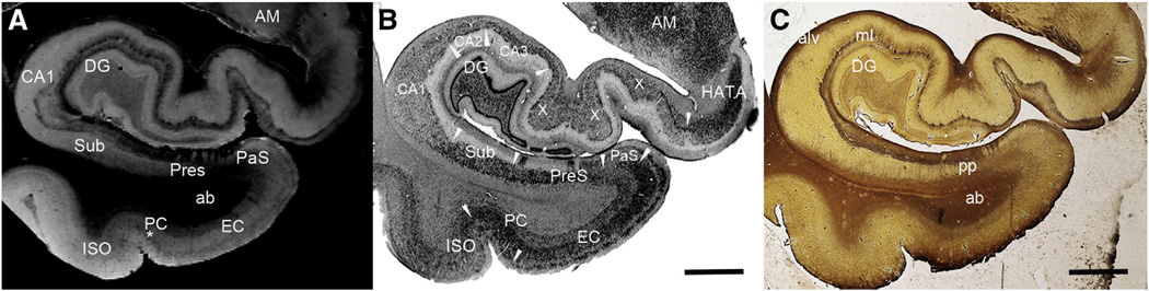
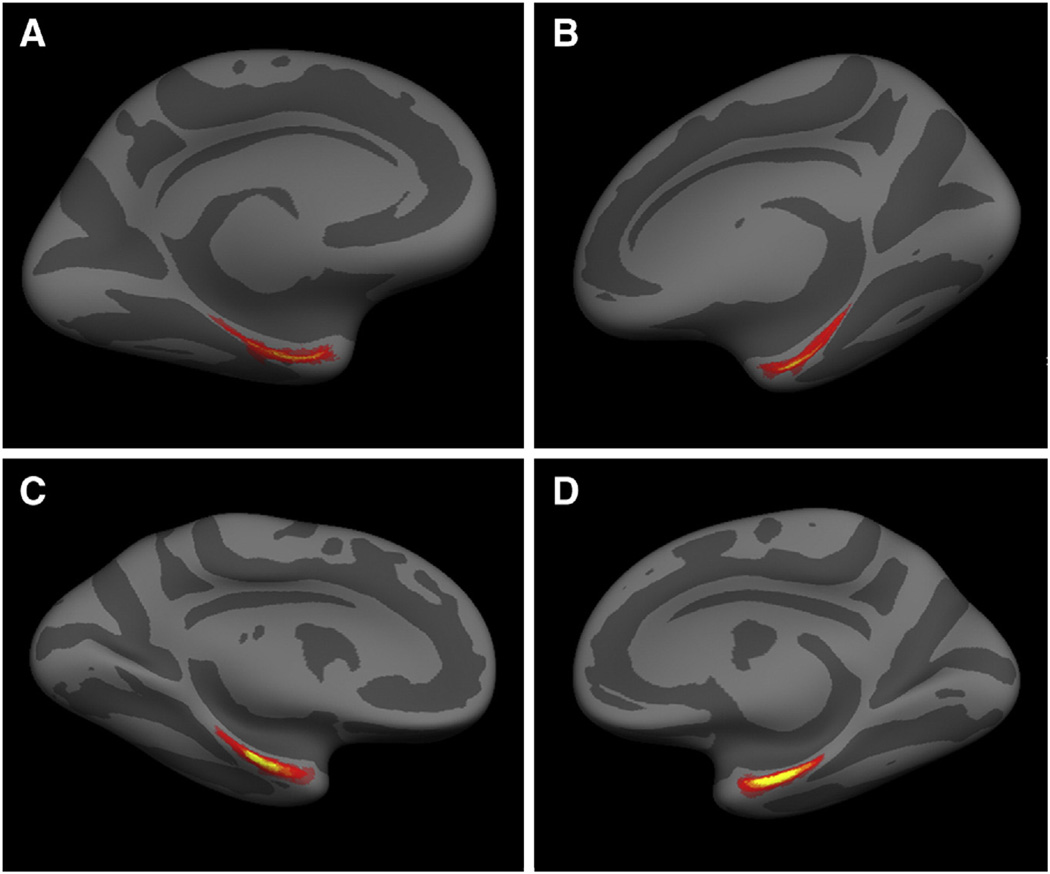
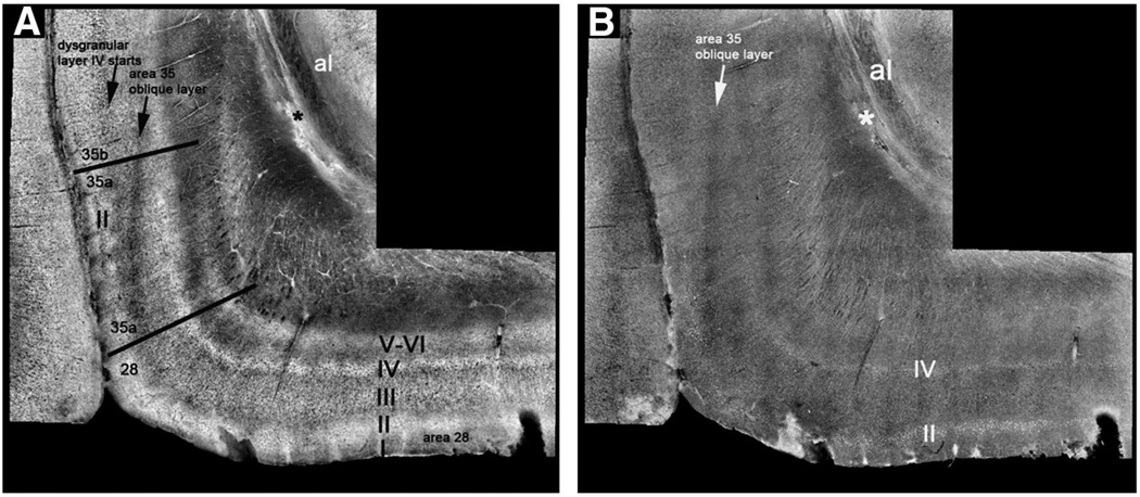
Recent advancements in radio frequency coils, field strength and sophisticated pulse sequences have propelled modern brain mapping and have made validation to biological standards - histology and pathology - possible. The medial temporal lobe has long been established as a pivotal brain region for connectivity, function and unique structure in the human brain, and reveals disconnection in mild Alzheimer's disease. Specific brain mapping of mesocortical areas affected with neurofibrillary tangle pathology early in disease progression provides not only an accurate description for location of these areas but also supplies spherical coordinates that allow comparison between other ex vivo cases and larger in vivo datasets. We have identified several cytoarchitectonic features in the medial temporal lobe with high resolution ex vivo MRI, including gray matter structures such as the entorhinal layer II 'islands', perirhinal layer II-III columns, presubicular 'clouds', granule cell layer of the dentate gyrus as well as lamina of the hippocampus. Localization of Brodmann areas 28 and 35 (entorhinal and perirhinal, respectively) demonstrates MRI based area boundaries validated with multiple methods and histological stains. Based on our findings, both myelin and Nissl staining relate to contrast in ex vivo MRI. Precise brain mapping serves to create modern atlases for cortical areas, allowing accurate localization with important applications to detecting early disease processes.
Keywords: Brodmann's area 28; Brodmann's area 35; Entorhinal; Localization; Mapping; Perirhinal.
Copyright © 2013 Elsevier Inc. All rights reserved.
Conflict of interest statement
The authors have no conflict of interest to disclose regarding this work.
Figures
References
-
- Amunts K, Zilles K. Advances in cytoarchitectonic mapping of the human cerebral cortex. Neuroimaging Clin. N. Am. 2001;11:151–169. (vii) - PubMed
-
- Amunts K, Kedo O, Kindler M, Pieperhoff P, Mohlberg H, Shah NJ, Habel U, Schneider F, Zilles K. Cytoarchitectonic mapping of the human amygdala, hippocampal region and entorhinal cortex: intersubject variability and probability maps. Anat. Embryol. (Berl.) 2005;210:343–352. - PubMed
-
- Arnold SE, Hyman BT, Flory J, Damasio AR, Van Hoesen GW. The topographical and neuroanatomical distribution of neurofibrillary tangles and neuritic plaques in the cerebral cortex of patients with Alzheimer's disease. Cereb. Cortex. 1991;1:103–116. - PubMed
-
- Arnold SE, Hyman BT, Van Hoesen GW, Damasio AR. Some cytoarchitectural abnormalities of the entorhinal cortex in schizophrenia. Arch. Gen. Psychiatry. 1991;48:625–632. - PubMed
-
- Arriagada PV, Growdon JH, Hedley-Whyte ET, Hyman BT. Neurofibrillary tangles but not senile plaques parallel duration and severity of Alzheimer's disease. Neurology. 1992;42:631–639. - PubMed
Publication types
MeSH terms
Grants and funding
- U24 RR021382/RR/NCRR NIH HHS/United States
- R01EB006758/EB/NIBIB NIH HHS/United States
- R01 NS052585-01/NS/NINDS NIH HHS/United States
- S10 RR019307/RR/NCRR NIH HHS/United States
- P41 RR014075/RR/NCRR NIH HHS/United States
- R01 NS052585/NS/NINDS NIH HHS/United States
- AG022381/AG/NIA NIH HHS/United States
- R01 AG008122/AG/NIA NIH HHS/United States
- S10 RR023043/RR/NCRR NIH HHS/United States
- RC1 AT005728-01/AT/NCCIH NIH HHS/United States
- R01 EB006758/EB/NIBIB NIH HHS/United States
- R01 AG022381/AG/NIA NIH HHS/United States
- U01 MH093765/MH/NIMH NIH HHS/United States
- R01 NS070963/NS/NINDS NIH HHS/United States
- P41-RR14075/RR/NCRR NIH HHS/United States
- 1S10RR019307/RR/NCRR NIH HHS/United States
- 1S10RR023043/RR/NCRR NIH HHS/United States
- 5R01AG008122-22/AG/NIA NIH HHS/United States
- 1R21NS072652-01/NS/NINDS NIH HHS/United States
- RC1 AT005728/AT/NCCIH NIH HHS/United States
- 1R01NS070963/NS/NINDS NIH HHS/United States
- 1S10RR023401/RR/NCRR NIH HHS/United States
- K01 AG028521/AG/NIA NIH HHS/United States
- 5U01-MH093765/MH/NIMH NIH HHS/United States
- R21 NS072652/NS/NINDS NIH HHS/United States
- S10 RR023401/RR/NCRR NIH HHS/United States
- K01AG028521/AG/NIA NIH HHS/United States
LinkOut - more resources
Full Text Sources
Other Literature Sources
Medical
