

Striatum-based circuitry of adolescent depression and anhedonia
- PMID: 23702452
- PMCID: PMC3762469
- DOI: 10.1016/j.jaac.2013.04.003
Striatum-based circuitry of adolescent depression and anhedonia
Abstract
Objective: Striatum-based circuits have been implicated in both major depressive disorder (MDD) and anhedonia, a symptom that reflects deficits of reward processing. Yet adolescents with MDD often exhibit a wide range of anhedonia severity. Addressing this clinical phenomenon, we aimed to use intrinsic functional connectivity (iFC) to study striatum-based circuitry in relation to categorical diagnosis of MDD and anhedonia severity.
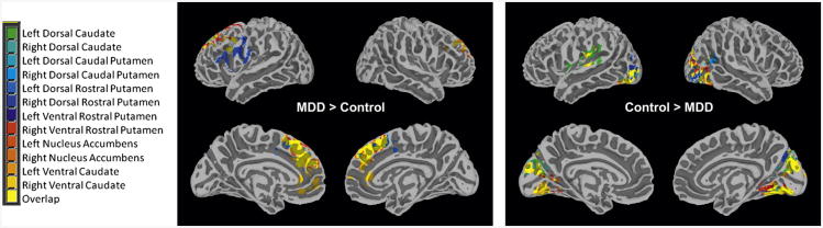
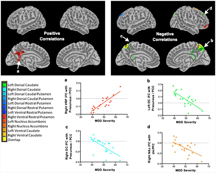
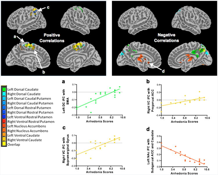
Method: A total of 21 psychotropic medication-free adolescents with MDD and 21 healthy controls (HC), group-matched for age and sex, underwent resting-state functional magnetic resonance imagining (fMRI) scans. Voxelwise maps indicating correlation strengths of spontaneous blood-oxygenation-level-dependent (BOLD) signals among 6 bilateral striatal seeds (dorsal caudate, ventral caudate, nucleus accumbens, dorsal-rostral putamen, dorsal-caudal putamen, ventral-rostral putamen) and the remaining brain regions were compared between groups. Relationships between striatal iFC and severity of MDD and anhedonia were examined in the MDD group. Analyses were corrected for multiple comparisons.
Results: Adolescents with MDD manifested increased iFC between all striatal regions bilaterally and the dorsomedial prefrontal cortex (dmPFC), as well as between the right ventral caudate and the anterior cingulate cortex (ACC). MDD severity was associated with iFC between the striatum and midline structures including the precuneus, posterior cingulate cortex, and dmPFC. However, distinct striatal iFC patterns involving the pregenual ACC, subgenual ACC, supplementary motor area, and supramarginal gyrus were associated with anhedonia severity.
Conclusions: Although MDD diagnosis and severity were related to striatal networks involving midline cortical structures, distinct circuits within the reward system were associated with anhedonia. Findings support the incorporation of both categorical and dimensional approaches in neuropsychiatric research.
Copyright © 2013 American Academy of Child and Adolescent Psychiatry. Published by Elsevier Inc. All rights reserved.
Figures
References
-
- Yorbik O, Birmaher B, Axelson D, Williamson DE, Ryan ND. Clinical characteristics of depressive symptoms in children and adolescents with major depressive disorder. J Clin Psychiatry. 2004;65:1654–1659. quiz 1760-1651. - PubMed
-
- Ryan ND, Puig-Antich J, Ambrosini P, et al. The clinical picture of major depression in children and adolescents. Arch Gen Psychiatry. 1987;44:854–861. - PubMed
Publication types
MeSH terms
Grants and funding
LinkOut - more resources
Full Text Sources
Other Literature Sources
