. 2013 Jun 14;435(4):733-9.
doi: 10.1016/j.bbrc.2013.05.052. Epub 2013 May 20.

Chondrogenic capacity and alterations in hyaluronan synthesis of cultured human osteoarthritic chondrocytes

Affiliations

Chondrogenic capacity and alterations in hyaluronan synthesis of cultured human osteoarthritic chondrocytes

Yohei Ono et al. Biochem Biophys Res Commun. .

Abstract

During osteoarthritis there is a disruption and loss of the extracellular matrix of joint cartilage, composed primarily of type II collagen, aggrecan and hyaluronan. In young patients, autologous chondrocyte implantation can be used to repair cartilage defects. However, for more elderly patients with osteoarthritis, such a repair approach is contraindicated because the procedure requires a large expansion of autologous chondrocytes in vitro leading a rapid, perhaps irreversible, loss of the chondrocyte phenotype. This study investigates whether osteoarthritic chondrocytes obtained from older patients can be expanded in vitro and moreover, induced to re-activate their chondrocyte phenotype. A decrease in chondrocyte phenotype markers, collagen II, aggrecan and SOX9 mRNA was observed with successive expansion of cells in monolayer culture. However, chondrogenic induction in three-dimensional pellet culture successfully rescued the expression of all three marker genes to native levels, even with 4th passage cells-cells representing an approximate 625-fold expansion in cell number. This data supports the use of osteoarthritic cells for autologous implantation repair. In addition, another set of gene products were explored as useful markers of the chondrocyte phenotype. Differentiated primary chondrocytes exhibited a common pattern of hyaluronan synthase isoforms that changed upon cell expansion in vitro and, reverted back to the original pattern following pellet culture. Moreover, the change in isoform pattern correlated with changes in the molecular size of synthesized hyaluronan.


Figures

Fig. 1
Fig. 1. Expression of chondrogenic marker genes and glycosaminoglycan release by human OA chondrocytes
The mRNA expression of COL2 (A), ACAN (B) and SOX9 (C) was quantified from total RNA isolated from native intact OA cartilage (white bars), primary chondrocytes (light grey bars) or passaged monolayers (dark grey bars) as well as P1–P4 re-differentiated in pellet cultures (gold bars). Samples were derived from OA patient material (n = 20, #P < 0.05; ##P < 0.01 Monolayer versus Pellet, *P < 0.05; **P < 0.01 versus Native Cartilage). (D) Glycosaminoglycan released into the medium of primary, P1–P3 cultures was analyzed by DMMB assay (n = 7, *P < 0.05 versus Primary). Values are mean ± SD (error bars). (E) Microscopic images of primary, P1 cells and P2 cells sub-cultured in alginate beads (R2).
Fig. 2
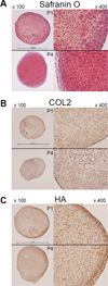
Fig. 2. Histological analysis of the pellet neocartilages from human OA chondrocytes
Representative sections of pellet neocartilages were stained with (A) Safranin O, (B) Immunohistochemistry staining for COL2 and, (C) HABP probe staining for HA. Comparisons were made between pellet-recovered P1 and P4 cultures isolated from an identical patient.
Fig. 3
Fig. 3. Expression of HAS isoforms and HA release by human OA chondrocytes
The mRNA expression of HAS1 (A), HAS2 (B) and HAS3 (C) was quantified from total RNA isolated from native intact OA cartilage (white bars), primary chondrocytes (light grey bars) or passaged monolayers (dark grey bars) as well as P1–P4 re-differentiated in pellet cultures (gold bars). Samples were derived from OA patient material (n = 20, #P < 0.05; ##P < 0.01 Monolayer versus Pellet, *P < 0.05; **P < 0.01 versus Native Cartilage). (D) HA released into the medium of primary, P1–P3 cultures was analyzed by HA ELISA (n = 7, n.s. = not significant). Values are mean ± SD (error bars).
Fig. 4
Fig. 4. Changes in HAS isoform expression pattern and the size determination of HA
(A) Summary of HAS isoform mRNA expression data taken from Fig. 3 for native intact OA cartilage and primary chondrocytes (left panel), monolayers (middle panel) and re-differentiated cells in pellet cultures (right panel). (B) Concentrated media from primary chondrocytes and P1–P4 expanded monolayers, derived from an identical patient, were run on agarose gels and stained for HA to determine the size distribution. (C) The same assay was performed using the media from the chondrocytes of another patient to determine the effects of human HAS1 or HAS2 overexpression on P1 monolayers, or sub-culture of P2 cells in alginate beads (R2). HA standards shown in the left hand lanes were from a commercial source (Lifecore Biomedical, Chaska, MN) with sizes ranging from 1,200–1,800 kDa, 180–350 kDa, and < 5 kDa (termed high, middle, and low molecular weight (HMW, MMW, and LMW)-HA, respectively).

Similar articles

Cited by

References

    1. Knudson CB, Knudson W. Hyaluronan and CD44: modulators of chondrocyte metabolism. Clin Orthop Relat Res. 2004;427:S152–S162. - PubMed
    1. Knudson W, Loeser RF. CD44 and integrin matrix receptors participate in cartilage homeostasis. Cell Mol Life Sci. 2002;59:36–44. - PMC - PubMed
    1. Nishida Y, Knudson CB, Nietfeld JJ, et al. Antisense inhibition of hyaluronan synthase-2 in human articular chondrocytes inhibits proteoglycan retention and matrix assembly. J Biol Chem. 1999;274:21893–21899. - PubMed
    1. Takahashi N, Knudson CB, Thankamony S, et al. Induction of CD44 cleavage in articular chondrocytes. Arthritis Rheum. 2010;62:1338–1348. - PMC - PubMed
    1. Akiyama H, Chaboissier MC, Martin JF, et al. The transcription factor Sox9 has essential roles in successive steps of the chondrocyte differentiation pathway and is required for expression of Sox5 and Sox6. Genes Dev. 2002;16:2813–2828. - PMC - PubMed

Publication types

Substances

LinkOut - more resources