. 2013 Aug;110(2):264-74.
doi: 10.1160/TH13-02-0135. Epub 2013 May 23.

Identification and characterisation of mutations associated with von Willebrand disease in a Turkish patient cohort

Affiliations

Identification and characterisation of mutations associated with von Willebrand disease in a Turkish patient cohort

Daniel J Hampshire et al. Thromb Haemost. 2013 Aug.

Abstract

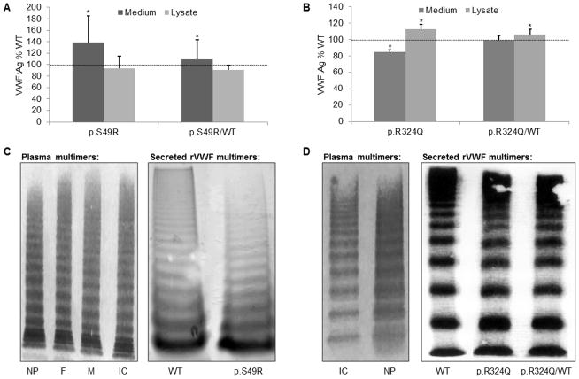
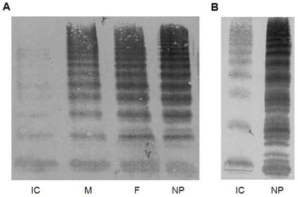
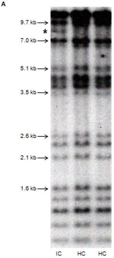
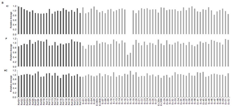
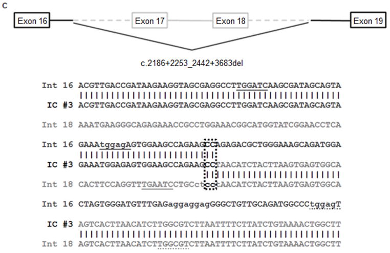
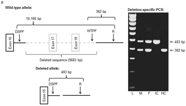
Several cohort studies have investigated the molecular basis of von Willebrand disease (VWD); however, these have mostly focused on European and North American populations. This study aimed to investigate mutation spectrum in 26 index cases (IC) from Turkey diagnosed with all three VWD types, the majority (73%) with parents who were knowingly related. IC were screened for mutations using multiplex ligation-dependent probe amplification and analysis of all von Willebrand factor gene (VWF) exons and exon/intron boundaries. Selected missense mutations were expressed in vitro. Candidate VWF mutations were identified in 25 of 26 IC and included propeptide missense mutations in four IC (two resulting in type 1 and two in recessive 2A), all influencing VWF expression in vitro. Four missense mutations, a nonsense mutation and a small in-frame insertion resulting in type 2A were also identified. Of 15 type 3 VWD IC, 13 were homozygous and two compound heterozygous for 14 candidate mutations predicted to result in lack of expression and two propeptide missense changes. Identification of intronic breakpoints of an exon 17-18 deletion suggested that the mutation resulted from non-homologous end joining. This study provides further insight into the pathogenesis of VWD in a population with a high degree of consanguineous partnerships.


Conflict of interest statement

Conflicts of interest

None declared.

Figures

Figure 1
Figure 1
Recombinant VWF (rVWF) expression in COS-7 cells (mean values from six independent experiments). A and B) Mutant rVWF (p.S49R and p.R324Q respectively) secreted into the culture medium and retained within the cell relative to WT rVWF only (100%). C and D) Comparison of patient multimer profile (electrophoresed on 2% SDS-agarose) and multimer profile of secreted rVWF (1.5% SDS-agarose) observed for p.S49R (rVWF in homozygous state only) and p.R324Q respectively (IC, index case; F, father; M, mother; NP, normal plasma; *p<0.05, two-tailed Student’s t-test).
Figure 2
Figure 2
Multimer profiles observed in IC #4 with 2A(IIC) VWD and their parents (M, mother: VWF:Ag 60 IU/dL, VWF:RCo 36 IU/dL, FVIII:C 98 IU/dL, no bleeding symptoms; F, father: VWF:Ag 26 IU/dL, VWF:RCo 30 IU/dL, FVIII:C 107 IU/dL, moderate bleeding symptoms) when electrophoresed on 2% (A) or 3% (B) SDS-agarose (NP, normal plasma).
Figure 3
Figure 3
Characterisation of the exon 17–18 deletion. A) Southern blot analysis of EcoRI digested DNA highlighting the novel ~8 kb fragment (*) observed in IC #3 but neither HC. B) Confirmation of the exon 17–18 deletion following dosage analysis by MLPA in the index case (IC), a heterozygous parent (P) and a healthy control (HC). C) Deletion breakpoints in intron 16 and intron 18 highlighting 2 bp microhomology (dotted border), deletion hotspot consensus sequences (solid underline), DNA polymerase arrest sites (lowercase) and DNA polymerase a/b frameshift hotspots (dotted underline) close to the breakpoint junctions. D) Multiplex PCR designed to detect the exon 17–18 deletion (DSPF, deletion-specific forward primer; WTPF, wild-type forward primer; R, reverse primer) further confirming the homozygous inheritance in the index case (IC) and heterozygous inheritance in both parents (F and M).
Figure 3
Figure 3
Characterisation of the exon 17–18 deletion. A) Southern blot analysis of EcoRI digested DNA highlighting the novel ~8 kb fragment (*) observed in IC #3 but neither HC. B) Confirmation of the exon 17–18 deletion following dosage analysis by MLPA in the index case (IC), a heterozygous parent (P) and a healthy control (HC). C) Deletion breakpoints in intron 16 and intron 18 highlighting 2 bp microhomology (dotted border), deletion hotspot consensus sequences (solid underline), DNA polymerase arrest sites (lowercase) and DNA polymerase a/b frameshift hotspots (dotted underline) close to the breakpoint junctions. D) Multiplex PCR designed to detect the exon 17–18 deletion (DSPF, deletion-specific forward primer; WTPF, wild-type forward primer; R, reverse primer) further confirming the homozygous inheritance in the index case (IC) and heterozygous inheritance in both parents (F and M).

References

    1. Sadler JE, Budde U, Eikenboom JCJ, et al. Update on the pathophysiology and classification of von Willebrand disease: a report of the Subcommittee on von Willebrand Factor. J Thromb Haemost. 2006;4:2103–14. - PubMed
    1. Rodeghiero F, Castaman G, Dini E. Epidemiological investigation of the prevalence of von Willebrand’s disease. Blood. 1987;69:454–9. - PubMed
    1. Werner EJ, Broxson EH, Tucker EL, et al. Prevalence of von Willebrand disease in children: a multiethnic study. J Pediatr. 1993;123:893–8. - PubMed
    1. Sadler JE. Von Willebrand disease type 1: a diagnosis in search of a disease. Blood. 2003;101:2089–93. - PubMed
    1. Gupta PK, Ahmed RPH, Sazawal S, et al. Relatively high frequency of VWD types 3 and 2 in a cohort of Indian patients: the role of multimeric analysis. J Thromb Haemost. 2005;3:1321–2. - PubMed

MeSH terms