

Combinatorial assessments of brain tissue metabolomics and histopathology in rodent models of human immunodeficiency virus infection
- PMID: 23702663
- PMCID: PMC3889226
- DOI: 10.1007/s11481-013-9461-9
Combinatorial assessments of brain tissue metabolomics and histopathology in rodent models of human immunodeficiency virus infection
Abstract
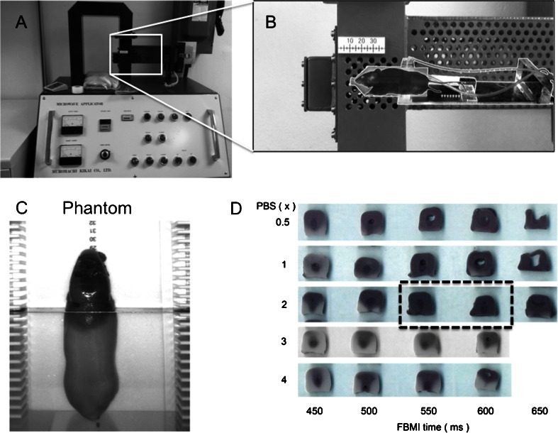
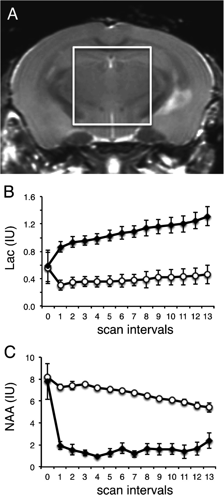
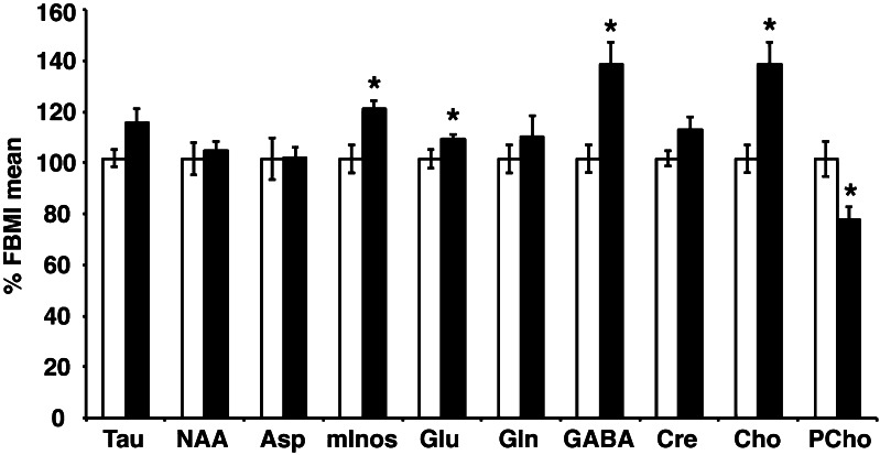
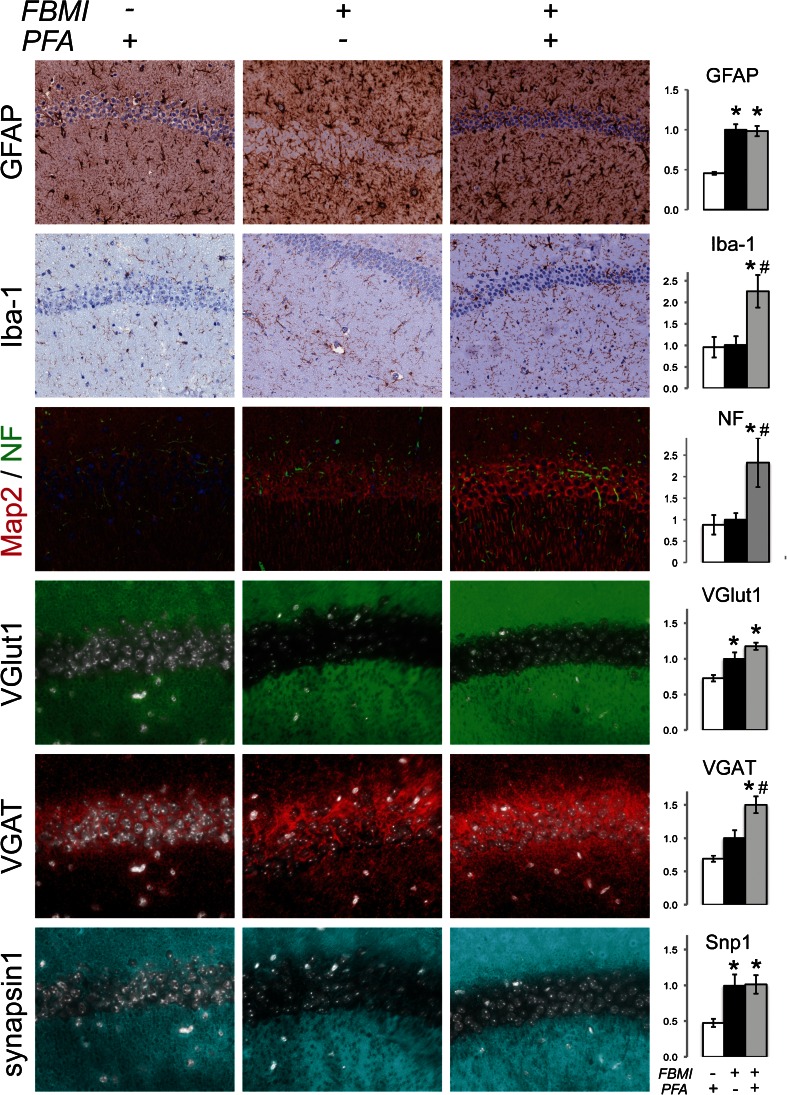
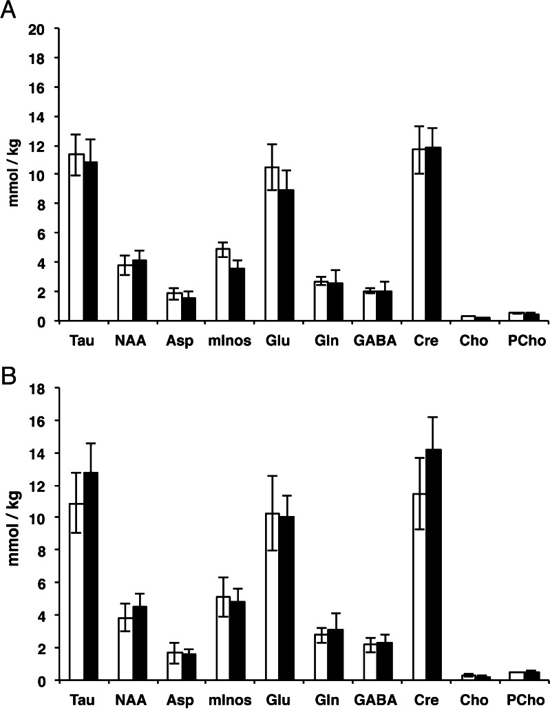
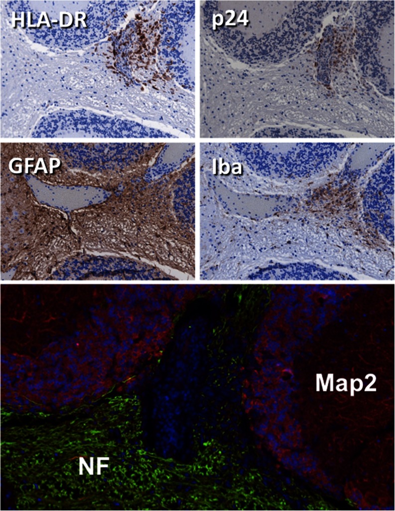
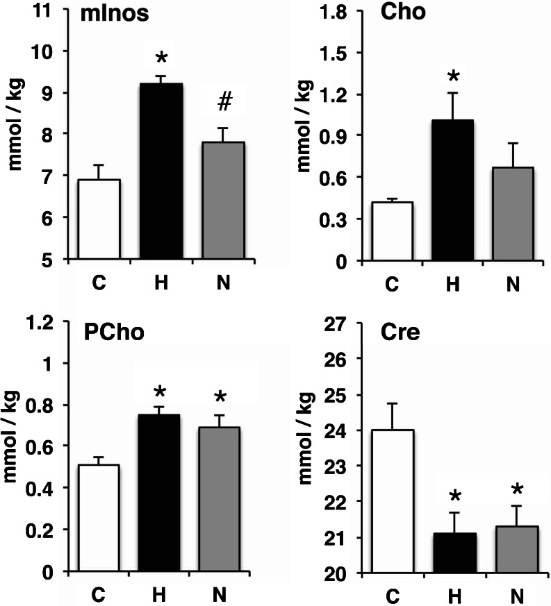
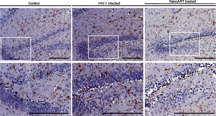
Metabolites are biomarkers for a broad range of central nervous system disorders serving as molecular drivers and byproducts of disease pathobiology. However, despite their importance, routine measures of brain tissue metabolomics are not readily available based on the requirements of rapid tissue preservation. They require preservation by microwave irradiation, rapid freezing or other methods designed to reduce post mortem metabolism. Our research on human immunodeficiency virus type one (HIV-1) infection has highlighted immediate needs to better link histology to neural metabolites. To this end, we investigated such needs in well-studied rodent models. First, the dynamics of brain metabolism during ex vivo tissue preparation was shown by proton magnetic resonance spectroscopy in normal mice. Second, tissue preservation methodologies were assessed using liquid chromatography tandem mass spectrometry and immunohistology to measure metabolites and neural antigens. Third, these methods were applied to two animal models. In the first, immunodeficient mice reconstituted with human peripheral blood lymphocytes then acutely infected with HIV-1. In the second, NOD scid IL2 receptor gamma chain knockout mice were humanized with CD34+ human hematopoietic stem cells and chronically infected with HIV-1. Replicate infected animals were treated with nanoformulated antiretroviral therapy (nanoART). Results from chronic infection showed that microgliosis was associated with increased myoinostitol, choline, phosphocholine concentrations and with decreased creatine concentrations. These changes were partially reversed with nanoART. Metabolite responses were contingent on the animal model. Taken together, these studies integrate brain metabolomics with histopathology towards uncovering putative biomarkers for neuroAIDS.
Figures
References
-
- Bathena SP, Huang J, Epstein AA, Gendelman HE, Boska MD, Alnouti Y. Rapid and reliable quantitation of amino acids and myo-inositol in mouse brain by high performance liquid chromatography and tandem mass spectrometry. J Chromatogr B Analyt Technol Biomed Life Sci. 2012;893–894:15–20. doi: 10.1016/j.jchromb.2012.01.035. - DOI - PMC - PubMed
-
- Bonneh-Barkay D, Bissel SJ, Wang G, Fish KN, Nicholl GC, Darko SW, Medina-Flores R, Murphey-Corb M, Rajakumar PA, Nyaundi J, Mellors JW, Bowser R, Wiley CA. YKL-40, a marker of simian immunodeficiency virus encephalitis, modulates the biological activity of basic fibroblast growth factor. Am J Pathol. 2008;173(1):130–143. doi: 10.2353/ajpath.2008.080045. - DOI - PMC - PubMed
MeSH terms
Grants and funding
LinkOut - more resources
Full Text Sources
Other Literature Sources
