Review
. 2013 Jun;13(6):397-411.
doi: 10.1038/nrc3526.

Neuroblastoma: developmental biology, cancer genomics and immunotherapy

Affiliations
Review

Neuroblastoma: developmental biology, cancer genomics and immunotherapy

Nai-Kong V Cheung et al. Nat Rev Cancer. 2013 Jun.

Abstract

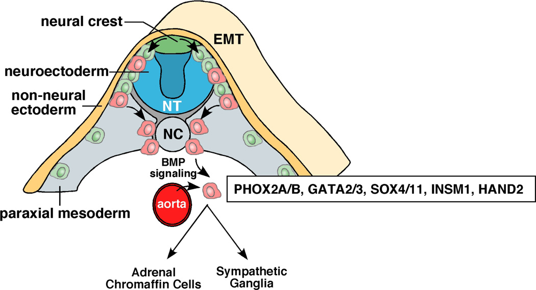
Neuroblastoma is a solid tumour that arises from the developing sympathetic nervous system. Over the past decade, our understanding of this disease has advanced tremendously. The future challenge is to apply the knowledge gained to developing risk-based therapies and, ultimately, improving outcome. In this Review we discuss the key discoveries in the developmental biology, molecular genetics and immunology of neuroblastoma, as well as new translational tools for bringing these promising scientific advances into the clinic.


Conflict of interest statement

Conflicts of Interest

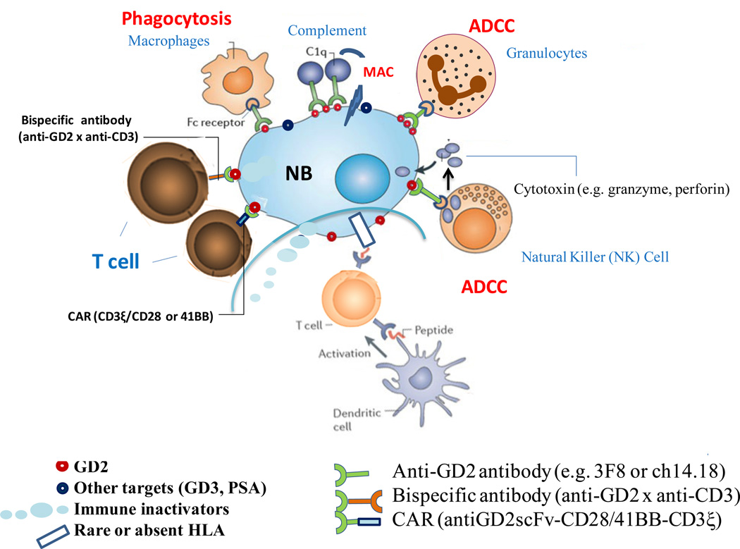
MSKCC has a patent application on hu3F8 and NKC was named as one of the inventors. MSKCC has licensed the patent on beta-glucan to Biotec Pharmacon, and the patent on antibody 8H9 to United Therapeutics, and NKC was named as one of the inventors for both agents. Clinical trials of hu3F8 are funded by the NCI (NKC) and the DOD (NKC).

Figures

Figure 1
Figure 1. Development of the sympathoadrenal lineage of the neural crest
As cells of the neural crest (green/red cells) migrate, they undergo epithelial-mesenchymal transition (EMT). A subset of cells (red) migrates toward the dorsal aorta as they commit to the sympathoadrenal lineage. This migration is directed in part by the expression of the chemokine receptor CXCR4 on the migrating neural crest progenitor cells (red) and the expression of the SDF-1 chemoattractant on the dorsal aorta. At the dorsal aorta, the migrating neural crest progenitor cells committed to the sympathoadrenal linege initiate their differentiation program in response to BMP signalling emanating from the dorsal aorta. A series of transcription factors including PHOX2A/B, ASCL1, GATA2/3, SOX4/11, INSM1 and HAND2 are upregulated. Shortly after these transcription factors are induced, neuronal markers are upregulated along with genes that encode enzymes required for catecholamine biosynthesis such as tyrosine hydroxylase (TH) and dopamine beta hydroxylase (DBH). From that point, the cells commit to the adrenal chromaffin lineage[G] or become sympathetic ganglia. Abbreviations: NT, neural tube; NC, notochord; EMT, epithelial to mesenchymal transition.
Figure 2
Figure 2. ALK and ATRX mutations in NB
A | Schematic representation of ALK protein structure and mutations found in NB. The low-density lipoprotein domain, two MAM domains, and the transmembrane and kinase domains of ALK are shown. R1275, F1174 and F1245 are three most common ALK mutations in neuroblastoma; the frequency of these mutations is provided in parenthesis. Other low frequency mutations are denoted with an asterisk. The tyrosine kinase inhibitor crizotinib is in clinical trials and other second-generation ALK inhibitors are in development. (Reproduced with permission from Carpenter et al). B | In a recent WGS study of stage-4 NBs from different age groups, ATRX mutations were identified, including in-frame deletions, missense, nonsense and frame-shift single-nucleotide variations. Mutations in ATRX were significantly associated with age at diagnosis. Mutations in ATRX were mutually exclusive of MYCN amplification and were associated with alternative lengthening of telomeres (ALT). Importantly, previous studies have shown that ALT in NB is mutually exclusive of MYCN amplification. More common among older patients (Group 4, 1), ALT is associated with chronic disease and poor survival. The 5-year OS for patients with ALT-positive NB is 0%; that of patients with ALT-negative is 52%ATRX mutations may contribute to ALT in NB cells and are associated with poor OS among older patients. (Modified with permission from Cheung et al).
Figure 3
Figure 3. Immunotherapy of Neuroblastoma
NB evades T-cells by downregulating or losing HLA expression, thereby interfering with the afferent arm (priming through dendritic cells), homing of T-cells to NB, and the CTL effector phase of adaptive immunity. Soluble inhibitors of immune response (e.g. FasL, gangliosides) are constantly released into the tumor stroma to impair cellular immunity. In addition, NB recruits pro-tumor macrophages and silences NK cells. Myeloid suppressor cells and T-reg can also suppress immunity. The paucity of mutations in NB compared to adult cancers like melanoma, the immaturity of the immune system in young patients, their massive disease and the intensive chemotherapy all combine to make NB poorly immunogenic for T-cells. Carbohydrate differentiation antigens (e.g. GD2, GD3 and polysialic acid (PSA)), all of which being classically T-independent antigens, offered alternative targets for antibody-based therapies. In the presence of monoclonal antibodies (e.g. 3F8 or ch14.18) specific for GD2, NB loses their defense and becomes highly susceptible to (1) NK (natural killer) cell mediated antibody-dependent cell mediated cytotoxicity (ADCC), (2) granulocyte mediated ADCC, (3) complement mediated cytotoxicity by binding to C1q thereby activating the complement cascade, delivering membrane attack complex (MAC) to tumor cell membrane, and (4) monocyte-macrophage mediated cytotoxicity. Even polyclonal T-cells can be retargeted to kill NB through MAbs in the form of chimeric antigen receptors (CAR) or bispecific antibodies (anti-GD2 x anti-CD3). CARs are anti-tumor single chain Fv fragments (scFv) genetically fused through a transmembrane domain to T-cell activating motifs (CD3 ξ and CD28/41BB) and transfected into killer lymphocytes. (Modified with permission from Scott et al.)

Similar articles

Cited by

References

    1. Maris JM. Recent advances in neuroblastoma. N Engl J Med. 2010;362:2202–2211. - PMC - PubMed
    1. Brodeur GM. Neuroblastoma: biological insights into a clinical enigma. Nat Rev Cancer. 2003;3:203–216. - PubMed
    1. Cohn SL, et al. The International Neuroblastoma Risk Group (INRG) classification system: an INRG Task Force report. Journal of clinical oncology : official journal of the American Society of Clinical Oncology. 2009;27:289–297. - PMC - PubMed
    1. Pugh TJ, et al. The genetic landscape of high-risk neuroblastoma. Nat Genet. 2013 - PMC - PubMed
    1. Molenaar JJ, et al. Sequencing of neuroblastoma identifies chromothripsis and defects in neuritogenesis genes. Nature. 2012;483:589–593. - PubMed

Publication types